Clear Sky Science · en
A feasibility study of deep learning-based segmentation of the inferior alveolar nerve on magnetic resonance neurography
Why this tiny dental nerve matters
The nerve running inside your lower jaw helps you feel your teeth, lip, and chin. Dentists try hard not to damage it during procedures, yet it can be difficult to see clearly on standard scans. This study explores whether a special type of MRI plus artificial intelligence can outline that nerve more directly, which could one day make common dental surgeries safer and planning more precise.
Limits of current jaw imaging
Today, dentists usually rely on panoramic X rays and cone beam CT scans to estimate where this nerve lies. These images show the bony canal that surrounds the nerve, not the nerve itself, and the canal edges are clearly visible in only about six out of ten people. When a patient has numbness or pain after an implant, CT often cannot tell whether only the bony wall is broken or if the nerve inside is bruised or cut. This uncertainty makes it hard to judge how serious an injury is and what recovery to expect.
A scan that shows nerves directly
Magnetic resonance neurography is a refined form of MRI designed to highlight nerves by suppressing signals from blood vessels and reducing motion artifacts. In the face and jaw, it can reveal changes such as swelling or damage along nerve fibers, offering information that bone based scans cannot. Despite these strengths, it has rarely been used with automated tools that could quickly and consistently trace small nerves. The authors set out to see whether a deep learning system could learn to segment the inferior alveolar nerve directly on these nerve focused MRI images.
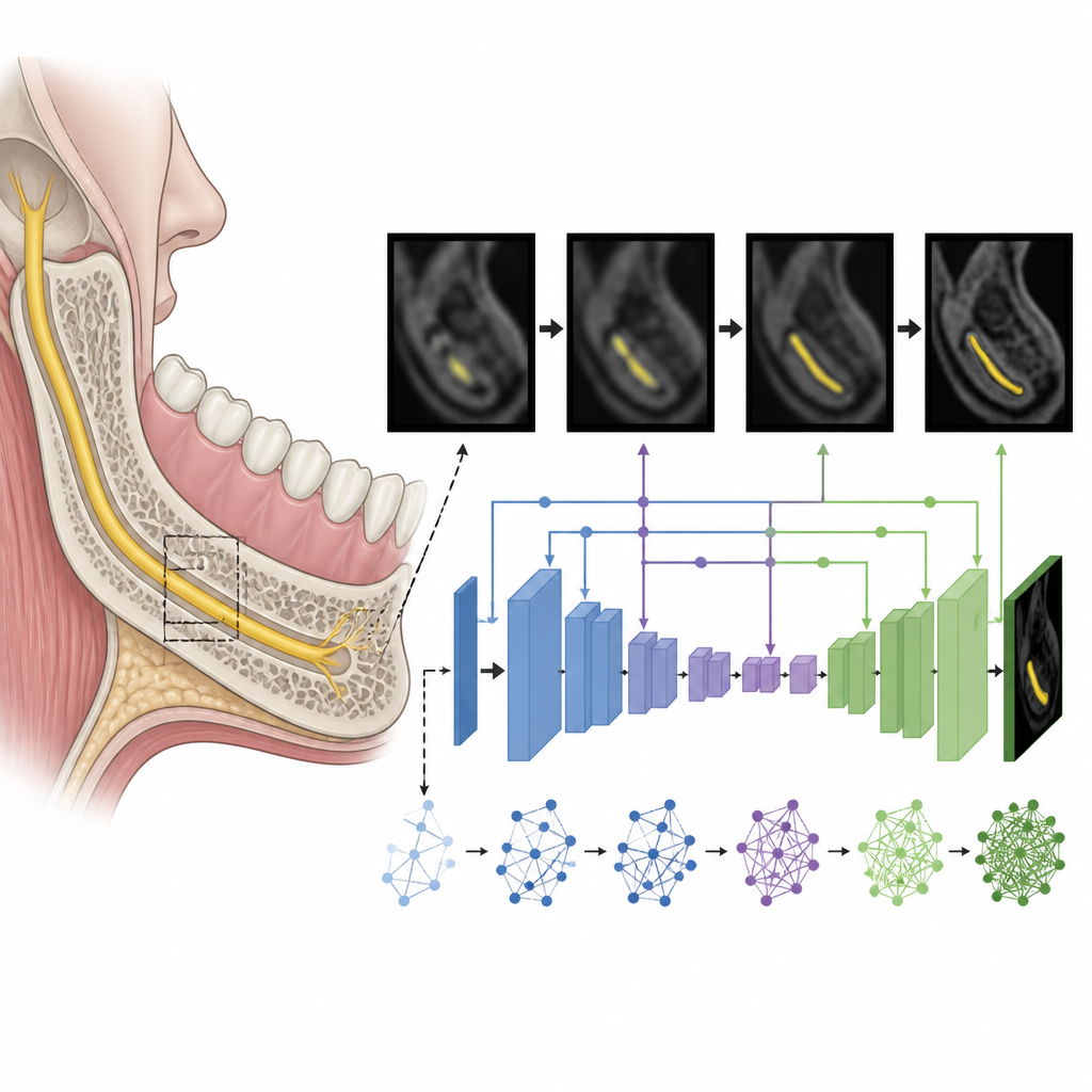
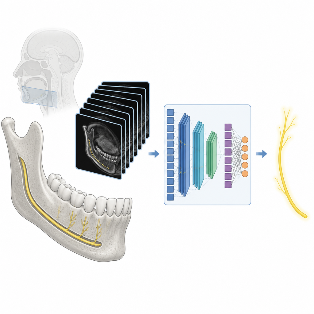
Teaching a computer to trace the nerve
The team gathered more than six thousand images from 51 healthy lower jaw nerves in 29 patients who had undergone magnetic resonance neurography for nerve problems. An expert radiologist carefully marked the nerve on each image slice to create a trusted reference. The researchers then preprocessed the images into uniform squares and trained a specialized neural network shaped to capture both fine detail and broader context, so it could pick out the thin, faint nerve against similar looking soft tissues. They compared this model with six leading segmentation networks that are widely used for small medical structures.
How well the model performed
The new system achieved higher accuracy than all six competing methods on four standard measures of segmentation quality. On average, its predicted nerve outlines overlapped the expert markings to a degree similar to what has been reported when two human readers segment other peripheral nerves. The researchers also looked beyond raw numbers at different kinds of failure: cases where the nerve was missed entirely, where the traced path jumped away from the true course, or where the borders were too loose or too tight. Their model had the lowest rate for each failure type. Even in many scans affected by bright streaks from dental metal, the system still managed to follow the nerve closely, although in one particularly distorted case every model failed.
What this could mean for dental care
This work does not yet deliver a push button tool for routine clinic use, and it was tested on a modest number of patients from a single center using normal nerves only. Still, it shows that nerve focused MRI combined with a tailored deep learning model can segment the inferior alveolar nerve with promising reliability. In the future, such tools could lighten the burden of manual tracing, give surgeons clearer maps of the nerve before placing implants or performing jaw surgery, and help track subtle changes in injured nerves over time.
Citation: Choi, Y.J., Han, S., Lee, C. et al. A feasibility study of deep learning-based segmentation of the inferior alveolar nerve on magnetic resonance neurography. Sci Rep 16, 15433 (2026). https://doi.org/10.1038/s41598-026-45392-6
Keywords: inferior alveolar nerve, dental imaging, magnetic resonance neurography, deep learning segmentation, oral surgery planning