Clear Sky Science · en
Metabolic changes in normal-appearing white matter associate with MRI measures of disease burden in relapsing-remitting multiple sclerosis over three years
Why hidden brain changes matter in multiple sclerosis
For people living with multiple sclerosis, or MS, standard brain scans often focus on the bright or dark spots that mark obvious areas of damage. Yet many patients continue to worsen even when these visible spots do not seem to change. This study asks a simple but important question: is there more going on in the brain’s wiring than regular scans can see, and can more sensitive imaging pick up early warning signs of silent disease activity?
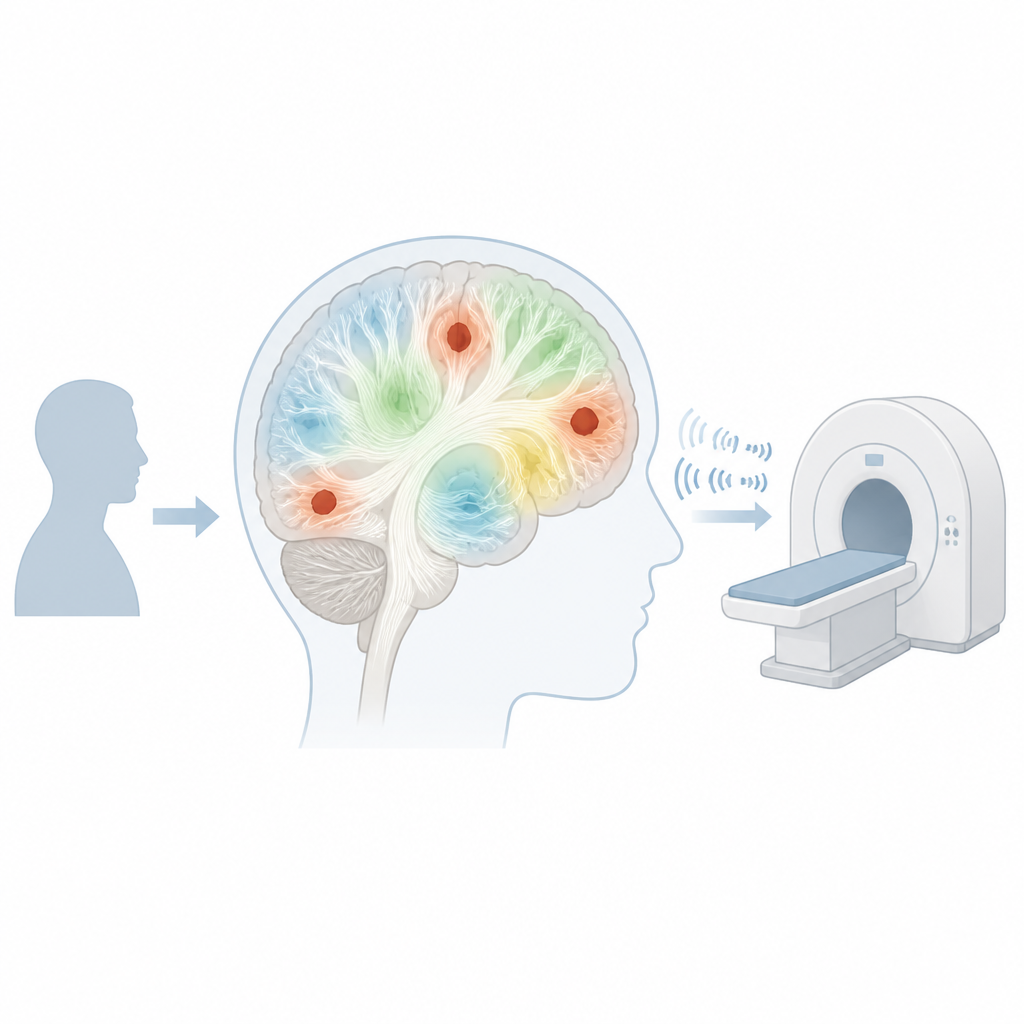
Looking beyond what standard scans can see
MS attacks the brain and spinal cord, damaging the insulated wiring that helps nerve cells talk to each other. On routine MRI scans, this damage shows up as lesions, but much of the surrounding white matter can still look normal. The researchers focused on this so-called normal-appearing white matter, asking whether its chemistry might already be altered in ways that reveal ongoing injury. To do this, they used a powerful 7-tesla MRI scanner equipped with a spectroscopic imaging technique that can measure tiny amounts of certain brain chemicals related to nerve cell health and support cell activity.
Tracking brain chemistry over three years
The team followed 20 people with relapsing–remitting MS and compared them with 20 healthy volunteers. Each participant with MS was scanned yearly, for up to three years, with a special two-dimensional spectroscopic scan placed over a region of white matter that often contains MS damage. The researchers focused on two key chemicals: total N-acetylaspartate, which reflects the health and function of nerve fibers, and myo-inositol, which is linked to support cells that become active in response to injury. By looking at ratios between these chemicals, they could infer whether nerve fibers were being lost, support cells were ramping up, or both.
Chemical signals of silent damage
Compared with healthy volunteers, people with MS already showed an imbalance in brain chemistry in white matter that still looked normal on standard scans. Their levels of the nerve-fiber marker were relatively lower, while the marker of support cell activity was higher when expressed as a ratio. Over the three-year follow-up, the ratio of myo-inositol to N-acetylaspartate continued to rise, suggesting that low-grade inflammatory and degenerative processes were quietly ongoing even when patients were largely stable in everyday function and did not show major loss of brain volume. These subtle changes could therefore be picking up damage earlier than traditional measures such as overall brain shrinkage.
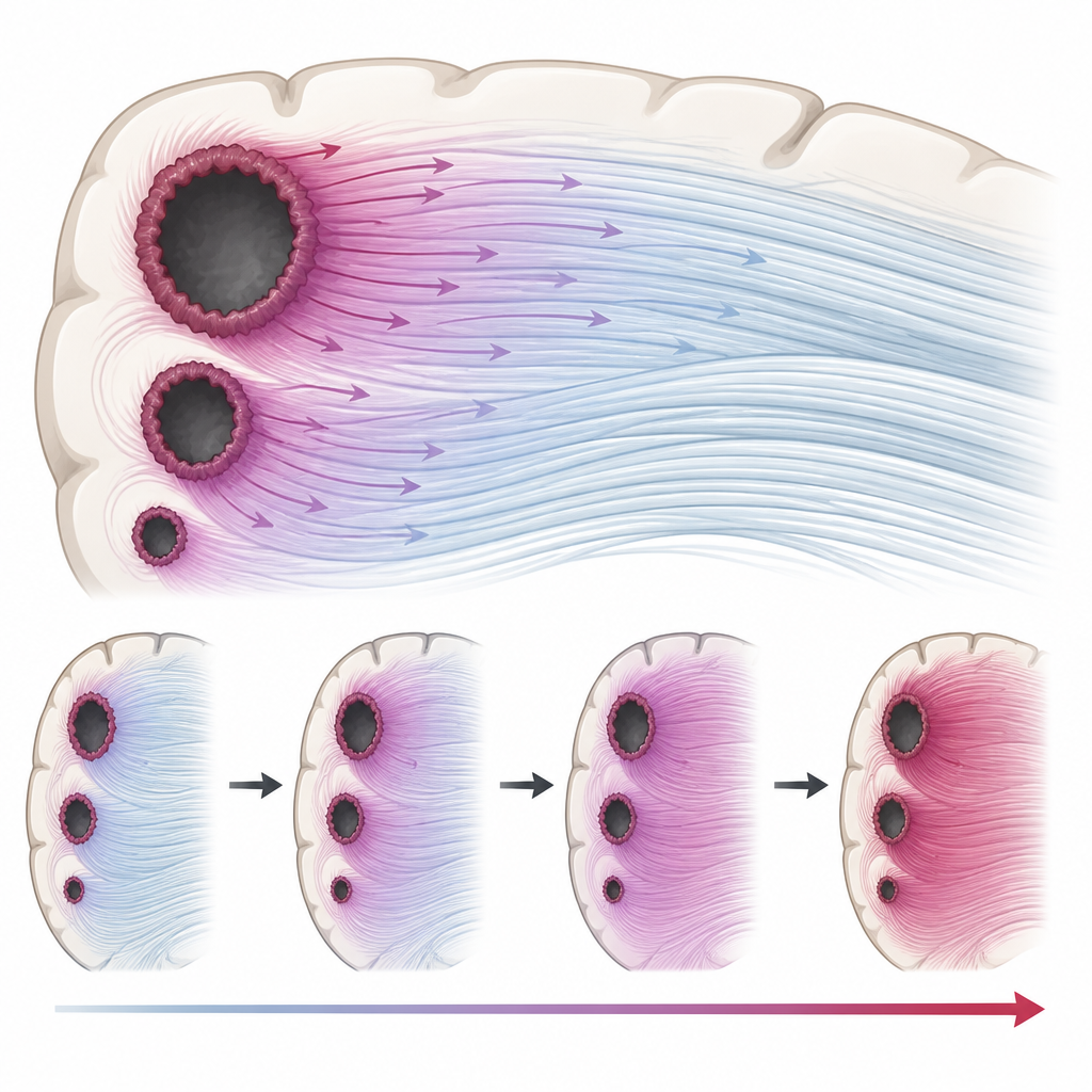
The special role of rim lesions
The study paid particular attention to a newer type of lesion called a paramagnetic rim lesion, which appears as a spot with a dark rim on certain high-field MRI scans and is thought to mark areas of chronic, smouldering inflammation. People with these rim lesions showed stronger signs of nerve fiber loss and more pronounced changes in the white matter chemistry around them. The more rim lesions a person had, the faster the myo-inositol to N-acetylaspartate ratio rose over time in nearby normal-appearing white matter. This pattern suggests that these chronic lesions act as long-term sources of damage that slowly affect surrounding wiring, even when no new attacks are seen.
What this means for people with MS
In simple terms, this work shows that the brain’s chemistry can reveal ongoing MS damage in areas that still look normal on conventional MRI. The persistent rise in the chemical ratio linked to support cell activation and nerve fiber stress, especially in people with rim lesions, points to slow, widespread injury that standard scans may miss. While the study is small and exploratory, it supports the idea that advanced spectroscopic imaging at very high magnetic fields could become a valuable tool for detecting silent disease progression earlier, refining risk assessment, and one day helping doctors tailor treatment before visible damage and disability accumulate.
Citation: Zöchner, A., Bogner, W., Dal-Bianco, A. et al. Metabolic changes in normal-appearing white matter associate with MRI measures of disease burden in relapsing-remitting multiple sclerosis over three years. Sci Rep 16, 14808 (2026). https://doi.org/10.1038/s41598-026-45342-2
Keywords: multiple sclerosis, white matter, brain imaging, paramagnetic rim lesions, magnetic resonance spectroscopy