Clear Sky Science · en
Structure and function of mouse lens suture examined by 2-photon fluorescence microscopic imaging
Why the eye’s tiny seams matter
When you look at the world, a clear eye lens quietly bends light so that images land in sharp focus on the retina. This paper explores what happens when the hidden seams inside the lens, called sutures, lose their orderly structure in mice. By using advanced, gentle microscopy that can see inside a living eye, the researchers reveal how disturbed sutures and damaged lens fibers are linked with cataracts, the clouding of the lens that often blurs vision with age.
The lens seen from the inside
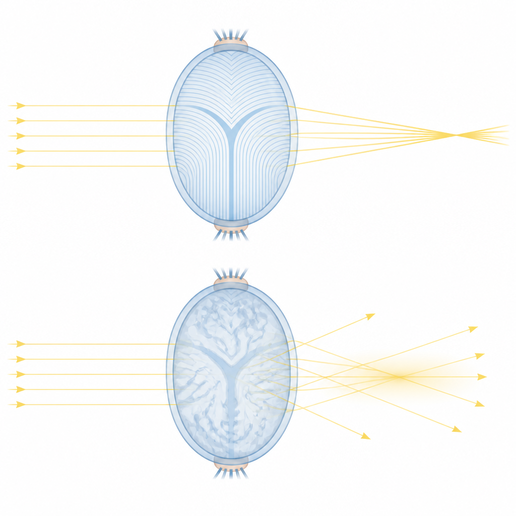
The lens is built from long, transparent cells stacked like layers of an onion. At the front and back poles of the lens, the tips of these cells meet and form branching patterns that look like the letter Y when viewed from above. These sutures help the lens keep its shape, stay mechanically sturdy, and maintain the even internal structure needed for clear vision. For years, scientists suspected that sutures might also act as tiny channels for nutrients and fluids, but most studies relied on dissected or fixed lenses, which can disturb delicate three dimensional organization.
Looking into living mouse lenses
To see sutures in their natural state, the team used two photon fluorescence microscopy, a type of laser imaging that can build detailed three dimensional pictures deep inside living tissue with minimal damage. They compared normal mice with mice lacking a protein called KLPH, which is known to be important for normal suture formation and which develop cataract like clouding. In normal lenses, the familiar Y and double Y suture patterns appeared in an orderly, predictable way at different depths. In contrast, lenses without KLPH showed a jumble of Y, double Y, and star like patterns that shifted more randomly with depth, along with poor alignment between the front and back sutures.
Holes, bubbles, and hidden debris
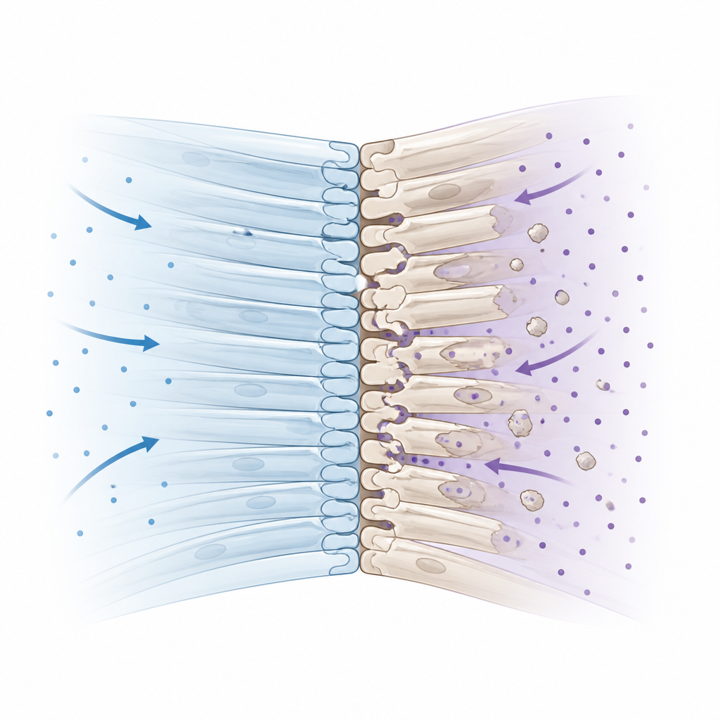
Imaging also uncovered voids and bubble like structures near the suture junctions in both normal and mutant lenses. To find out whether these spaces were filled with fluid between cells or were inside the cells themselves, the researchers soaked freshly removed lenses in a fluorescent dye that cannot cross intact cell membranes. The dye spread through the spaces between healthy lens fibers but did not strongly enter the central void at the suture junctions, suggesting this region is made of tightly packed cell ends with very little room between them. In the mutant lenses, many of the enlarged vacuoles and central voids showed reduced dye and cell membrane signals, and some contained small ring like or amorphous bits that likely represent leftover membrane fragments from damaged fibers.
How damaged seams may cloud vision
When the team measured how much light related signal passed through the lenses during imaging, they found that signal faded more quickly in mutant lenses than in normal ones, consistent with poorer transparency. The disorganized, more complex suture layouts and the presence of fiber end remnants and extra voids would be expected to bend and scatter light instead of letting it pass smoothly. This supports the idea that the KLPH protein is needed for lens fiber tips to join cleanly at the sutures, creating a stable interface that keeps the lens as a single, well organized unit. Without KLPH, fiber ends fail to integrate properly, leading to structural weak points and clutter that interfere with clear light transmission.
What this means for cataracts
For non specialists, the key message is that cataracts are not just a simple clouding of an otherwise uniform lens. They can arise from subtle breakdowns at the microscopic seams where lens cells meet. This study shows that in a mouse model of cataract, these seams become irregular and littered with cellular debris, and that the sutures do not act as open fluid channels as once thought. Instead, healthy sutures behave like sealed joints that help keep the lens solid and clear. By providing detailed, three dimensional markers of these changes in living eyes, the work lays a foundation for testing how future treatments might protect or restore lens structure before vision is lost.
Citation: Zhang, Q., Zhu, J., Painter, T. et al. Structure and function of mouse lens suture examined by 2-photon fluorescence microscopic imaging. Sci Rep 16, 14788 (2026). https://doi.org/10.1038/s41598-026-45299-2
Keywords: eye lens, cataract, lens sutures, two photon microscopy, mouse model