Clear Sky Science · en
CT-to-MRI translation of medical volume data based on an enhanced diffusion model
Why turning one scan into another matters
Hospitals often rely on several kinds of medical scans to see what is happening inside the body. A CT scan is quick and good at showing bones, while MRI is slower but reveals soft tissues such as the brain and organs in great detail. Getting both types can be costly, time consuming, and sometimes risky for patients. This study explores whether computers can learn to turn a three dimensional CT scan into an MRI like scan, so doctors can gain MRI style views even when only CT data are available.
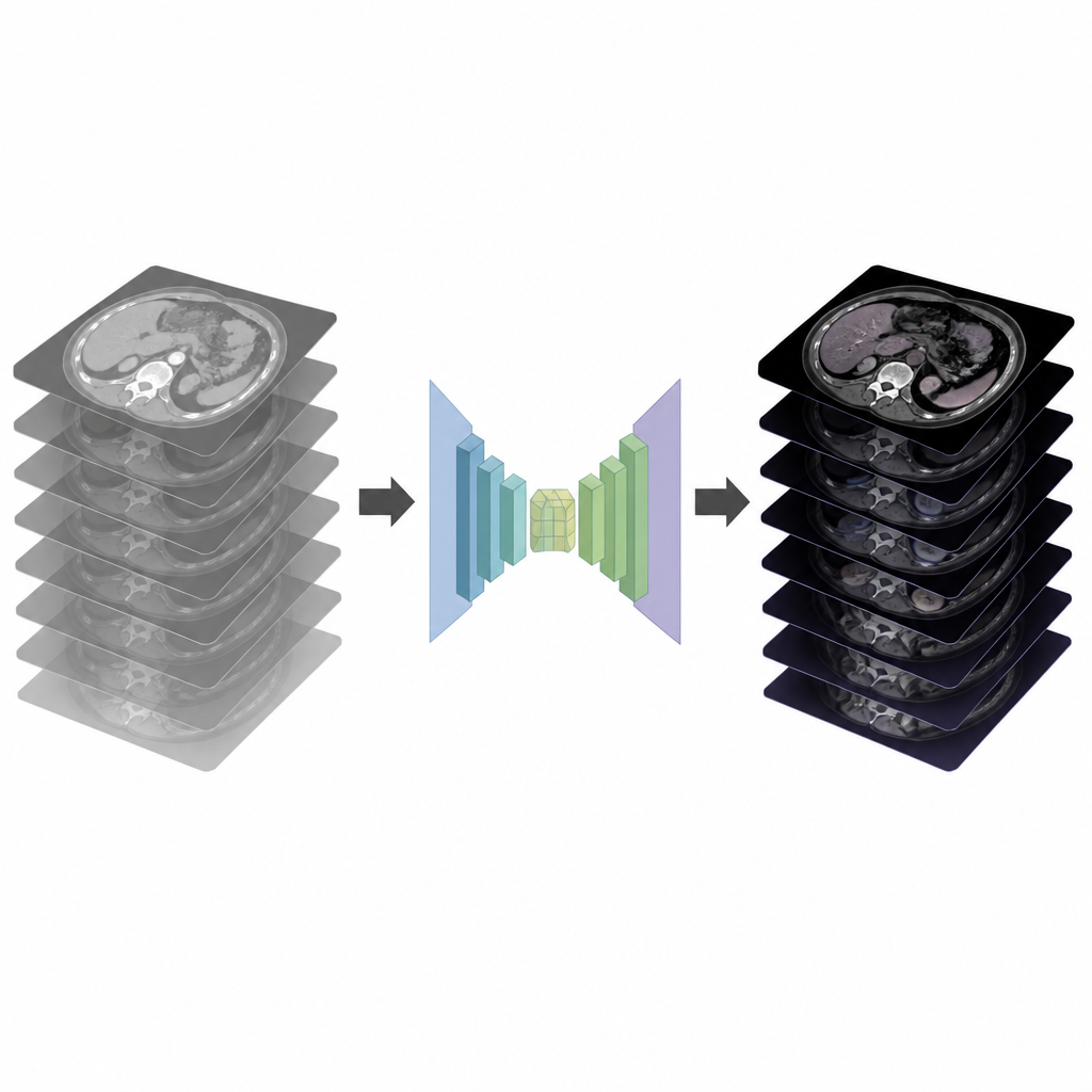
Different scans tell different stories
Each medical imaging method highlights different features of the body. CT is excellent for detecting fractures, bleeding, and lung problems, while MRI is preferred for examining the brain, spine, joints, and many internal organs. In an ideal world, every patient would receive all the scans needed, but MRI machines are expensive and often scarce, and long scan times can delay urgent treatment. Being able to generate MRI like images directly from existing CT data could give doctors extra information quickly, especially in emergencies or in hospitals with limited equipment.
From noisy patterns to clear pictures
The researchers focus on full three dimensional volumes rather than individual images. This is important because medical scans are stacks of slices, and their spatial relationships matter for surgery planning and tumor tracking. Converting entire volumes is harder than translating single pictures because the data are larger and the computer must keep details aligned across many slices. The team builds on a class of modern generative models called diffusion models, which learn to turn random noise into realistic images by stepwise denoising. They adapt this idea so that the model starts from noise and gradually produces an MRI like volume that is guided by the patient’s CT scan.
Smart building blocks for 3D medical data
To make diffusion work well on large medical volumes, the authors design a specialized network called enhanced DDPM. It uses two key building blocks. The first, a transform block, splits the flow of information into two paths to capture both fine details and broader shapes, helping preserve subtle structures. The second, a self attention less memory block, lets the model link distant parts of the volume so that the anatomy remains consistent, while using tricks like pooling and upsampling to keep memory use manageable on standard graphics cards. After the model generates an MRI like volume, a tailored post processing step performs global normalization and automatic contrast enhancement so that structures and potential lesions appear clearer and can be compared fairly across patients.
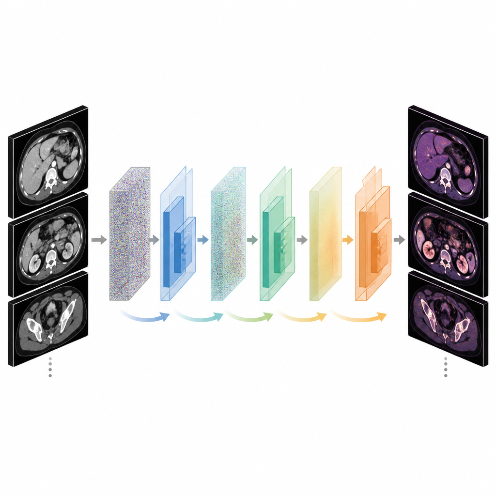
Testing on real brain and pelvis scans
The team trains and tests their method on a public collection of paired CT and MRI volumes from patients treated with radiotherapy to the brain or pelvis. Real clinical data rarely align perfectly across modalities, so some slices do not match exactly between CT and MRI. Despite about six percent of slices being imperfectly paired, the method still learns to produce realistic MRI like volumes. The authors compare their results with several leading techniques based on generative adversarial networks and standard diffusion models. Using quality measures that better reflect human visual judgment, such as structural similarity and feature based scores, their approach consistently yields images that more closely resemble the true MRI scans for both brain and pelvis regions.
What this means for future patient care
The study shows that an advanced diffusion based model can turn three dimensional CT scans into MRI like volumes that match real MRI data more closely than several existing methods. This could one day help doctors gain soft tissue views when MRI is unavailable or too slow, for example in emergency rooms or in clinics with limited resources. The authors caution that these synthetic images can still contain artifacts and should not replace real MRI for diagnosis. Instead, they may become a useful aid for planning and triage, especially once further research improves reliability, speeds up computation, and validates performance across more types of MRI scans and more diverse patient groups.
Citation: Ma, J., Chen, J. & Liang, A. CT-to-MRI translation of medical volume data based on an enhanced diffusion model. Sci Rep 16, 14774 (2026). https://doi.org/10.1038/s41598-026-45181-1
Keywords: CT-to-MRI translation, medical image synthesis, diffusion model, volumetric imaging, radiotherapy planning