Clear Sky Science · en
Neuroimaging insights from Wistar-Kyoto rats under chronic mild stress: morphological and metabolic brain correlates of treatment-resistant depression
Why this research matters
Many people with major depression do not get better, even after trying several standard antidepressant drugs. Doctors call this treatment-resistant depression, and it is especially disabling and difficult to study. This paper uses advanced brain imaging in a specialized rat model that mimics hard-to-treat depression to reveal how long-term stress reshapes the brain’s structure and chemistry, offering clues that could guide future therapies.
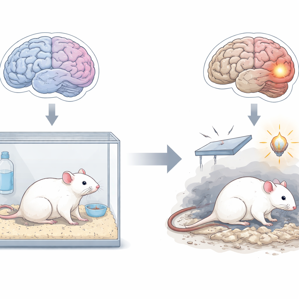
A rat stand-in for stubborn depression
Researchers focused on a strain of rat called Wistar-Kyoto, which naturally shows many depression-like traits: high stress sensitivity, low motivation, anxiety-like behavior and poor response to common antidepressants. They then added weeks of mild, unpredictable stressors—such as tilted cages, brief food or water deprivation and irregular lighting—to mimic the chronic stress many patients experience. This combined genetic and environmental vulnerability, known as the WKY/CMS model, closely resembles treatment-resistant depression because the animals do not improve with standard drugs but do respond to more intensive treatments like ketamine, brain stimulation and electroconvulsive therapy.
Seeing shape changes in the stressed brain
Using high-resolution magnetic resonance imaging, the team compared the brains of stressed Wistar-Kyoto rats with those of healthy Wistar control rats. The depressed-like rats had subtly shorter, more ellipsoid brains and noticeably enlarged fluid-filled spaces called ventricles. Two key regions involved in mood and memory—the cingulate part of the frontal cortex and the hippocampus—were thinner in the stressed animals. These patterns echo many MRI findings in people with major depression, where fronto-limbic areas often show loss of volume or thickness, especially in more severe or long-lasting illness.
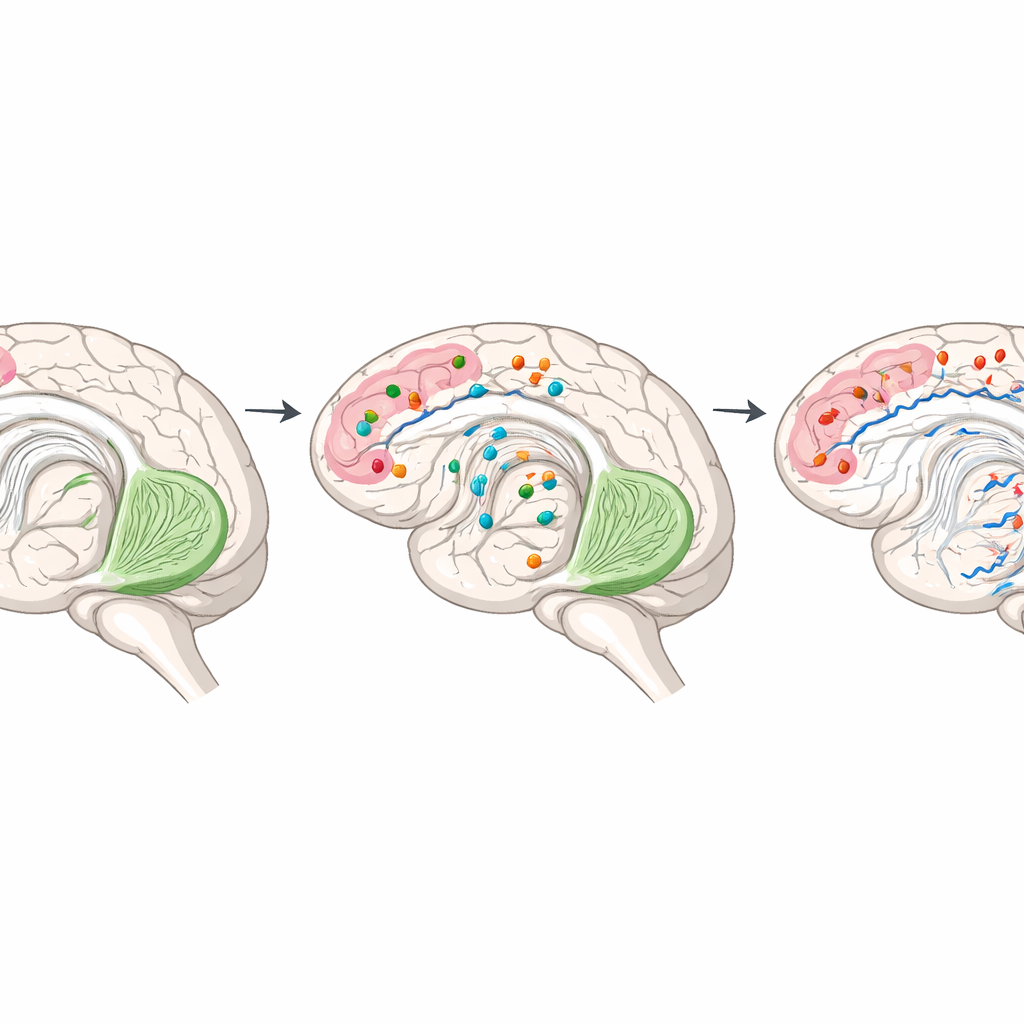
Chemical imbalances in mood and memory hubs
To look beyond shape and into chemistry, the researchers used proton magnetic resonance spectroscopy, a noninvasive method that estimates levels of naturally occurring brain chemicals in living tissue. In the prefrontal cortex of stressed rats, levels of glutamate and glutamine—key players in the brain’s main excitatory signaling system—were reduced, as was taurine, a molecule with protective and calming roles. At the same time, myo-inositol, often linked to glial cells and inflammatory processes, was increased. In the hippocampus, glutamine and choline-containing compounds, which support cell membranes and signaling, were lower, along with certain broad macromolecular signals thought to reflect amino-acid–rich proteins. Together, these shifts suggest disrupted communication between nerve cells and support cells and possible involvement of inflammation and altered energy use, themes that have also emerged in human depression studies.
Hidden wiring damage revealed by water movement
The team next examined the brain’s microscopic wiring with diffusion tensor imaging, which tracks how water moves along nerve fibers. In the stressed rats, water diffused more freely (higher mean diffusivity) in both the prefrontal cortex and hippocampus, and the directional organization of fibers (fractional anisotropy) was reduced in the hippocampus. These patterns are often interpreted as signs of weakened or thinned myelin sheaths, loss of axons or low-grade inflammation. Importantly, similar diffusion changes are repeatedly observed in people with treatment-resistant depression, particularly in the pathways that connect frontal regions with deeper emotional centers, and are thought to underlie problems with emotion regulation and cognition.
What this means for future treatments
Taken together, the findings show that rats modeling treatment-resistant depression have coordinated structural and chemical alterations in the same mood and memory circuits implicated in human illness. By extending this animal model to include detailed spectroscopy and diffusion measures, the study provides a set of measurable brain readouts that can be tracked as new treatments—such as advanced brain stimulation protocols or psychedelic-assisted therapies—are tested. For a layperson, the key message is that hard-to-treat depression is tied to real, measurable changes in brain wiring and chemistry, and this refined rat model gives scientists a powerful, ethically manageable way to probe those changes and evaluate therapies designed specifically for people who do not respond to standard antidepressants.
Citation: Gianmauro, P., Valentina, Z., Marta, B. et al. Neuroimaging insights from Wistar-Kyoto rats under chronic mild stress: morphological and metabolic brain correlates of treatment-resistant depression. Sci Rep 16, 10868 (2026). https://doi.org/10.1038/s41598-026-45121-z
Keywords: treatment-resistant depression, chronic stress, brain imaging, glutamatergic system, white matter