Clear Sky Science · en
Deep learning–based quantitative CT assessment of interstitial lung abnormalities: prognostic risk thresholds in a health screening population
Why tiny lung changes matter
Most people who go for a chest CT scan during a health checkup feel perfectly well. Yet these images sometimes reveal faint, patchy changes in the lungs that are easy to overlook. This study asks a simple but important question: when do such small, hidden changes become a warning sign for serious problems years later, and can computer tools help doctors spot that danger more reliably?
Quiet marks that may signal future illness
The researchers focused on subtle findings called interstitial lung abnormalities, or ILAs. These are small cloudy or netlike areas on CT scans that can be an early sign of scarring in the lung tissue. Because they are mild and often appear in people without symptoms, ILAs are usually found by chance and may not draw much attention. Past work suggested that people with these changes can have worse long term health, but no one knew how much abnormal tissue is enough to raise concern, especially in generally healthy adults coming for routine screening.
Using smart software to measure hidden damage
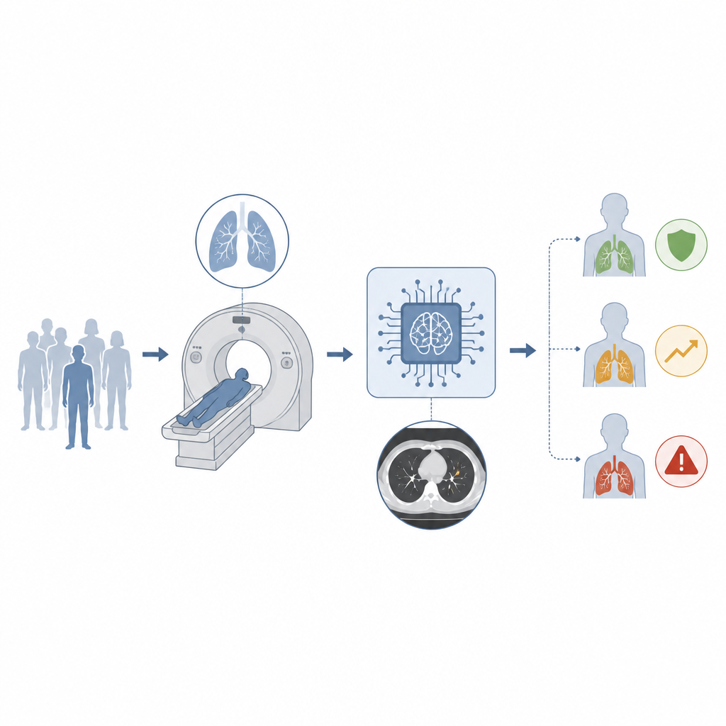
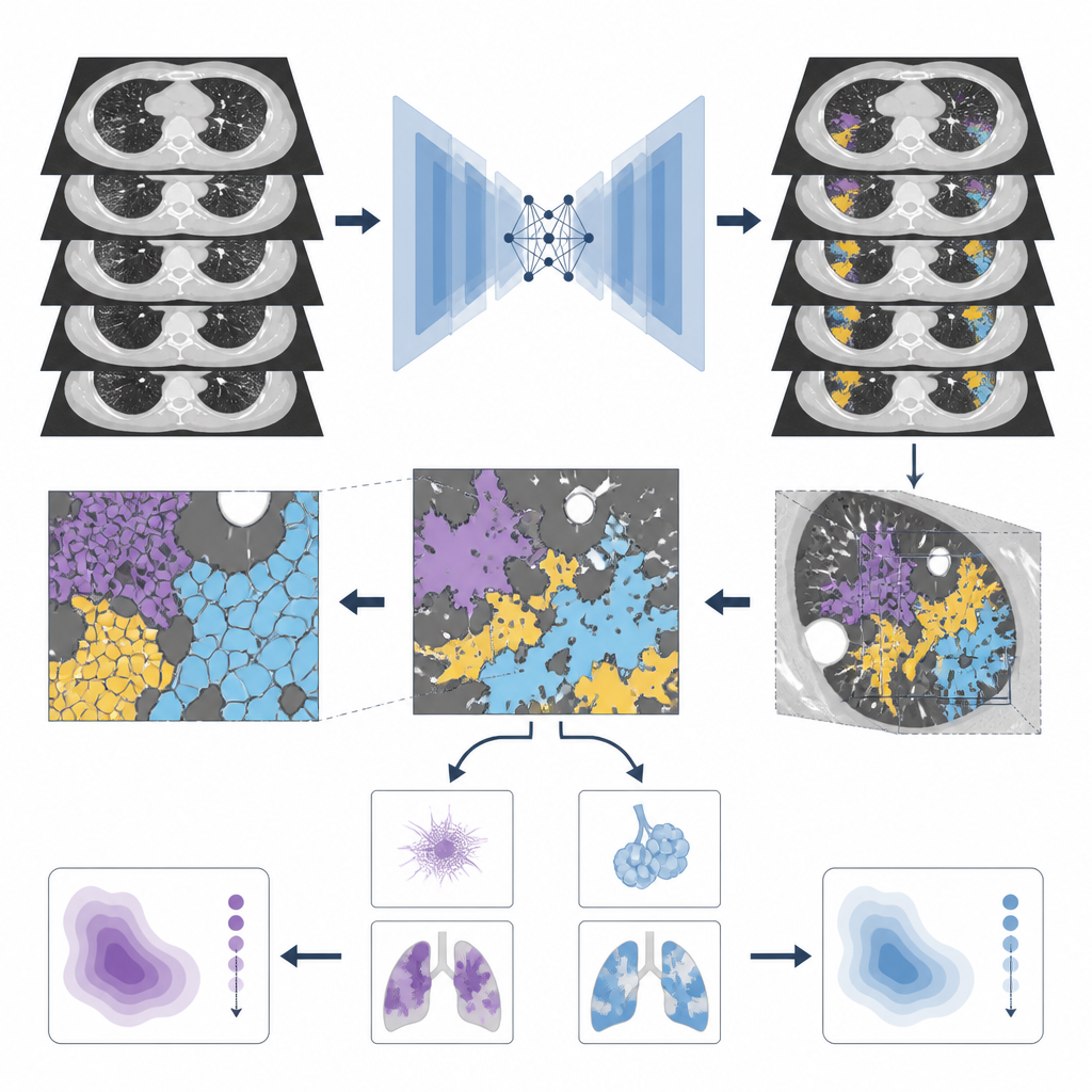
To tackle this, the team studied 3363 adults aged 50 and older who had chest CT scans as part of health screening at two hospitals between 2007 and 2013. Two experienced chest radiologists first reviewed each scan and sorted people into three groups: clear lungs, uncertain ILA, or definite ILA. The same scans were then analyzed by a deep learning program designed to pick out and measure abnormal lung textures across the entire lung. The software separated less worrisome hazy areas from patterns suggesting scarring and calculated what fraction of total lung volume each type occupied.
Finding the tipping points for risk
The participants were followed for over 11 years, with records checked for later development of interstitial lung disease, lung cancer, and death from any cause. The researchers tested many possible cutoffs for how much abnormal lung was present and used statistical models to see which levels best separated higher risk from lower risk, while adjusting for age, sex, smoking, body weight, lung function, and lung size. They found that when total abnormal tissue reached about 3 percent of the lung, or when scarring like changes reached about 0.3 percent, people were much more likely to die during the follow up period. Those above the 3 percent total mark had more than five times the risk of death compared with those below, and those above the 0.3 percent scarring mark had nearly three times the risk.
Links to lung disease and cancer
The same computer measurements also flagged people at higher risk of developing full fledged interstitial lung disease or lung cancer. Even relatively small amounts of abnormal tissue were tied to sharply higher chances of a later diagnosis of lung scarring disorders or lung tumors. Interestingly, most deaths in the study were not from lung causes, which suggests that these lung changes may be a visible sign of more general aging or frailty rather than acting alone to cause harm.
What this means for health checks
For patients and clinicians, the main message is that tiny lung changes on a screening CT scan should not be brushed aside, especially when they quietly occupy a few percent of the lung. By using deep learning tools to measure these areas in a consistent way, doctors can identify people whose scans cross the 3 percent total and 0.3 percent scarring thresholds and may need closer follow up. While these numbers are not a diagnosis on their own, they provide practical guideposts for turning faint shadows on a scan into clearer signals about long term health.
Citation: Lee, J.E., Suh, Y.J., Kim, K. et al. Deep learning–based quantitative CT assessment of interstitial lung abnormalities: prognostic risk thresholds in a health screening population. Sci Rep 16, 14852 (2026). https://doi.org/10.1038/s41598-026-45108-w
Keywords: interstitial lung abnormalities, chest CT, deep learning, lung fibrosis, health screening