Clear Sky Science · en
Correlation between human expert macular fluid height assessment and fluid volume quantification in neovascular age-related macular degeneration
Why this matters for everyday sight
As people live longer, more of us face age-related eye diseases that can steal central vision and make reading, driving, or recognizing faces difficult. One of the most serious forms, called wet age-related macular degeneration, causes fluid to leak into the back of the eye. Doctors must judge how much fluid is present to decide when to give injections that help protect sight. This study asks a simple question with big consequences: do traditional expert eye readings match what modern artificial intelligence tools see when they measure that fluid in three dimensions?
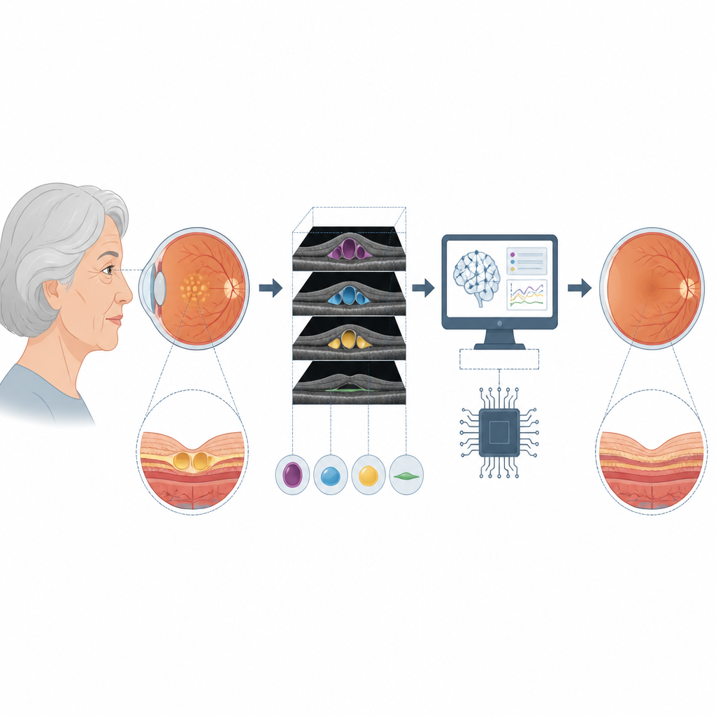
From flat snapshots to full volume views
Until recently, eye doctors and reading centers relied on thin cross-sectional images of the retina and measured the tallest point of fluid pockets on a single central slice. These "height" readings were used as stand-ins for overall disease activity and helped guide how often patients received drug injections into the eye. However, the retina is a three-dimensional tissue, and fluid can spread widely or cluster in small pockets far from the exact center. The researchers used an approved AI tool that examines the entire scan volume of the macula and calculates how much fluid is present in three key spaces within the retina, turning flat snapshots into detailed 3D maps.
Putting human judgment and AI side by side
The team analyzed eye scans from 890 people with untreated wet macular degeneration. Certified human readers measured the maximum height of three types of fluid within the central millimeter of the retina: fluid inside the retinal layers, fluid just under the light-sensing cells, and bulges where a supporting layer lifts up. The AI system examined the same scans, automatically outlining those fluid pockets pixel by pixel and calculating both their maximum height and their total volume within the center and across a wider six millimeter area. This direct comparison let the researchers test how closely expert measurements and AI calculations agreed.
Where the AI agreed and where it differed
For fluid inside the retina and for the raised areas under the pigment layer, AI height measurements were very close to the expert readings, showing strong agreement. For fluid sitting just under the light-sensitive cells, the match was more modest. In those cases, human readers tended to count slightly hazy material as part of the fluid, while the AI system left those regions out. When the researchers compared simple height at the center with the total amount of fluid in three dimensions, they found good links only within the narrow central zone. Once they looked at the wider six millimeter area, the connection weakened, especially for the shallow, spread-out subretinal fluid, showing that a single tallest point does not capture how much fluid truly fills the macula.
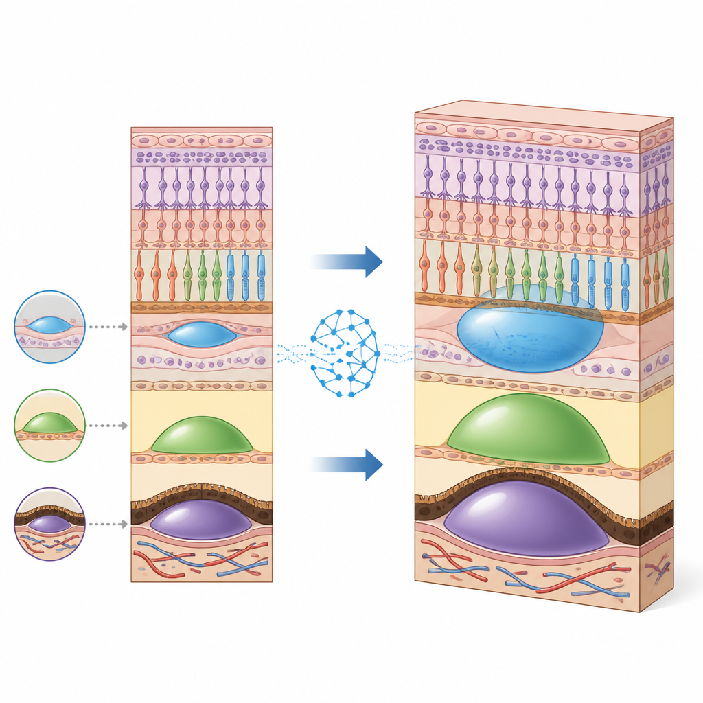
Seeing where fluid really builds up
By scanning the full macular area, the AI tool also revealed where fluid tends to peak. Fluid inside the retina most often reached its highest point near the very center, but almost as often just outside that zone. In contrast, subretinal fluid and the pigment layer bulges more commonly peaked in a ring around the center rather than directly beneath it. This pattern means that focusing only on the very center of the retina can miss important pockets of disease. Automated volume maps can highlight these off-center trouble spots quickly and consistently, without the time-consuming task of inspecting dozens of image slices by hand.
What this means for patients and clinicians
The study shows that for two major fluid types, AI-based measurements line up well with expert assessment, while also adding information that simple height readings cannot provide. It also makes clear that the tallest point of a fluid pocket is a poor stand-in for the full amount and spread of fluid across the macula. For patients, this suggests that AI tools may help eye specialists track disease activity more completely and tailor treatment schedules more closely to the real fluid burden in the eye. Rather than relying on a few manual measurements, doctors could use whole-volume, automated maps to decide when injections are truly needed, aiming for more precise and potentially more personalized care.
Citation: Steiner, S., Gerendas, B.S., Deak, G. et al. Correlation between human expert macular fluid height assessment and fluid volume quantification in neovascular age-related macular degeneration. Sci Rep 16, 14793 (2026). https://doi.org/10.1038/s41598-026-44982-8
Keywords: age-related macular degeneration, retinal fluid, optical coherence tomography, artificial intelligence, eye imaging