Clear Sky Science · en
A deep learning approach for the diagnosis and recurrence prediction of OKC
Why this matters for everyday health
Jaw cysts may sound rare, but they can quietly grow for years and return even after surgery, leading to repeated treatment and high medical costs. This study explores how artificial intelligence can help dentists and pathologists spot a particular jaw cyst called odontogenic keratocyst early and estimate the chance it will come back, with the goal of making care faster, more accurate, and easier to understand for both doctors and patients.
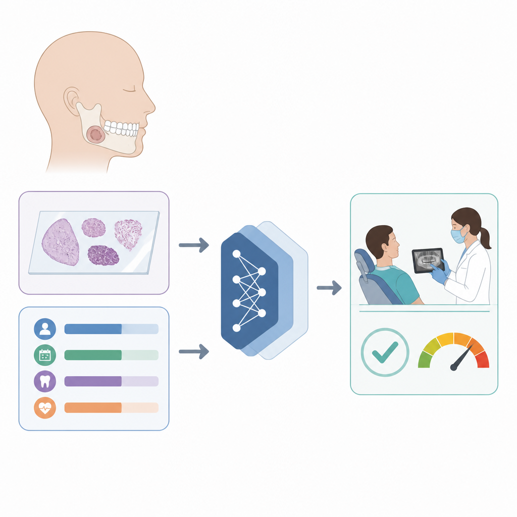
Understanding a stubborn jaw cyst
Odontogenic keratocyst is a benign tumor that develops in the bones of the jaw, most often in teenagers and young adults. It can be hard to diagnose because the tissue patterns under the microscope are complex and early symptoms are subtle. Even after surgery, the cyst often returns, which means patients may undergo repeated operations and long follow-up. Traditional diagnosis relies on experts carefully scanning huge digital microscope slides and piecing together scattered clinical details, a process that is time consuming and prone to differences in judgment between doctors.
Teaching computers to see and reason
The researchers built a computer system that learns from two kinds of information at once: digital tissue slides and basic clinical data such as age, how long symptoms have lasted, and where in the jaw the problem lies. First, the program automatically finds the useful tissue regions on each giant slide and cuts them into many smaller image patches. A deep learning network trained on natural images then turns each patch into a compact signature that captures fine details like the thickness of the keratin layer and the arrangement of cells. At the same time, another part of the system converts the clinical numbers and categories into a richer representation that can be combined with the image information.
Blending signals and showing its work
Rather than simply stacking image and clinical data side by side, the system uses an attention mechanism that learns how much weight to give each patch and each clinical factor for every patient. When the pictures are unclear, the model can lean more on the clinical clues, and when tissue patterns are striking, it can focus there. To make this process less of a black box, the authors added tools that highlight which parts of the slide most influenced the decision and which clinical values mattered most. Heat maps on the tissue images show red zones where the algorithm sees typical cyst features, while color coding on the data path reveals which patient factors tipped the balance toward or away from a diagnosis.
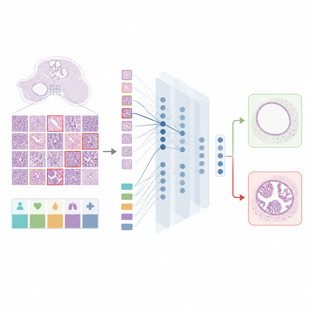
Looking ahead to the risk of coming back
The team also wanted to help doctors answer a question that patients care about deeply: Will this cyst return after surgery? For this, they designed a second model that focuses on follow-up records, including age, sex, exact location in the jaw, and how long patients were tracked after treatment. This model again uses attention to emphasize more informative factors, and a special loss function to cope with the fact that only a small fraction of patients experience recurrence. To open the model’s reasoning to inspection, the authors used a method that estimates how much each feature pushed the predicted risk up or down across many patients, revealing patterns such as certain jaw regions being linked to higher risk and longer follow-up without problems being linked to lower risk.
From research tool to clinic helper
In tests on real patient data from a major dental hospital, the combined system was more accurate at both diagnosing the cyst and predicting its return than several leading image and data analysis methods. Just as importantly, the authors wrapped these models into an information platform that allows clinicians to upload slides, enter basic clinical details, and receive not only a diagnosis and risk score but also visual explanations. While the study is based on data from a single center and still needs broader trials, it shows how carefully designed and interpretable AI could become a practical assistant in dental clinics, helping tailor follow-up plans and reducing the burden of repeated disease for young patients.
Citation: Chen, W., Qian, M., Zhang, M. et al. A deep learning approach for the diagnosis and recurrence prediction of OKC. Sci Rep 16, 14790 (2026). https://doi.org/10.1038/s41598-026-44979-3
Keywords: odontogenic keratocyst, jaw cyst, deep learning, recurrence prediction, digital pathology