Clear Sky Science · en
Deep learning approach to super-resolution correction of brain MRI motion artifacts for accurate hippocampal volumetry
Why clearer brain scans matter
As populations age, more people are living with memory problems and dementia. Doctors often rely on MRI scans of the brain to spot early changes in a small, deep structure called the hippocampus, which is crucial for forming new memories. But MRI scans take time, and even a simple sneeze or a brief touch to the face can blur the images, making the hippocampus look smaller or larger than it really is. This study explores whether an advanced form of artificial intelligence can "clean up" these motion-blurred brain scans, helping doctors measure the hippocampus more accurately without repeating long, uncomfortable exams.
Blurry pictures and memory loss
In conditions such as Alzheimer’s disease, the hippocampus slowly shrinks years before symptoms become obvious. Tracking its size over time can help doctors diagnose disease earlier and follow its progression. Unfortunately, MRI is very sensitive to motion: a short movement during a several-minute scan can create streaks and ghosting, similar to a wobbly long-exposure photograph. These artifacts can trick computer programs into overestimating or underestimating hippocampal volume, potentially pushing a person across a diagnostic threshold. Reducing this motion-related distortion is therefore key to trustworthy, quantitative brain measurements.
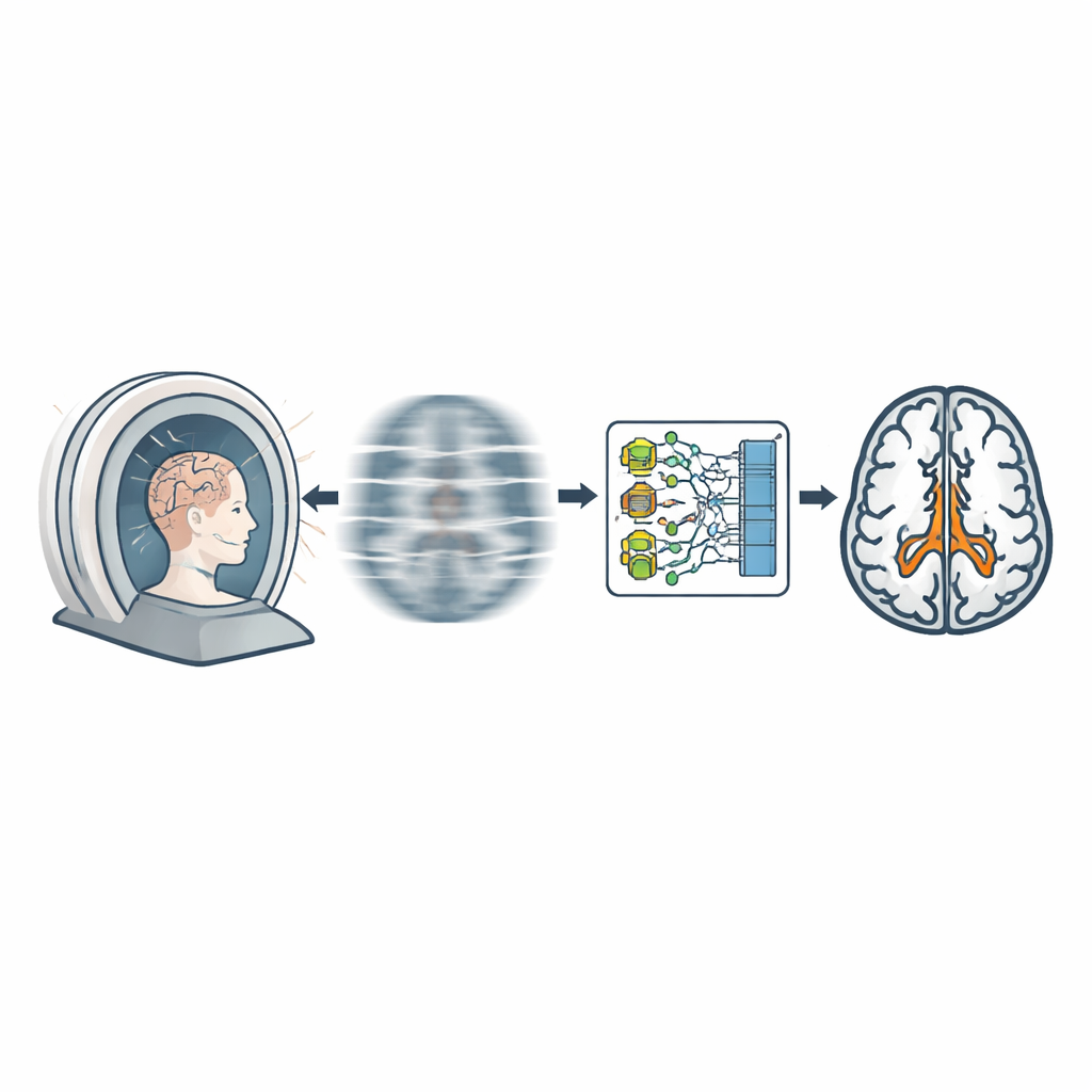
Teaching a computer to sharpen scans
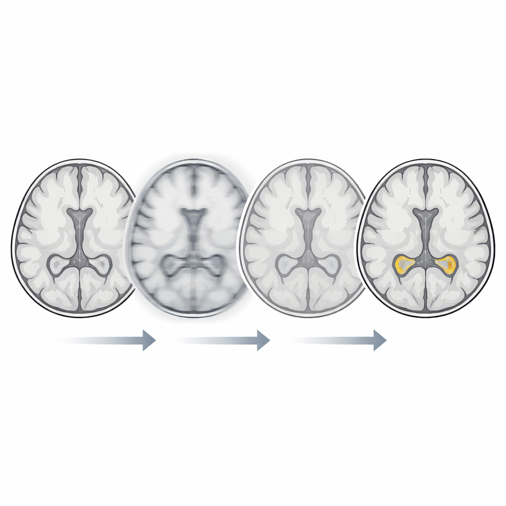
The researchers focused on a deep learning method called an enhanced deep super-resolution network, or EDSR. Super-resolution algorithms are designed to take a lower-quality image and reconstruct a sharper, more detailed version. Here, the team trained EDSR on thousands of high-quality brain scans from older adults, teaching it how fine brain structures should look. They then designed a two-step process for motion-blurred scans: first, the blurred images were gently downsampled, which softens the high-frequency distortions introduced by motion; second, EDSR rebuilt a high-resolution image from this smoothed version, aiming to restore true anatomy while suppressing artifacts.
Putting the method to the test
To see how well this approach worked, the researchers scanned 24 healthy young adults three times: once while keeping perfectly still, and twice while deliberately moving in realistic ways—sneezing or briefly touching their face—during the scan. The motion-free image served as the reference. The motion-corrupted images were then processed by EDSR to produce motion-corrected versions. An automated tool measured hippocampal volume from all three sets of images, and the team compared how far the motion and corrected measurements strayed from the true reference volume. They also calculated common image-quality scores that capture how similar each scan is to the original in terms of both pixel values and overall structure.
What improved and what stayed the same
The AI-based method clearly improved how the images looked on paper: across both sneeze-like and face-touch movements, the corrected scans had higher similarity scores and lower error measures than the uncorrected scans when compared to the motion-free reference. In other words, EDSR produced images that were consistently closer to what the scanner would have seen if the person had not moved. When it came to the actual hippocampal volume numbers, the story was more nuanced. In most cases, overall error did not change dramatically, but in the subgroup with the strongest motion during sneeze-type scans, the left hippocampus measurements became meaningfully more accurate after correction. In low-motion cases, EDSR usually had only a small effect, and in one subgroup it even slightly increased error on the right side, suggesting that indiscriminate use on nearly clean scans could be counterproductive.
Looking ahead to better brain care
Overall, the study shows that deep learning can make motion-blurred MRI scans look more like motion-free images and, in some high-motion situations, bring hippocampal measurements closer to their true values. While this is not yet a complete fix—some motion errors remain, and the test volunteers were healthy young adults—the work points toward a future where smart reconstruction tools quietly repair imperfect scans in the background. If refined and validated in older patients with real-world movement, such technology could reduce the need for repeat scans, shorten exam times, and make early dementia assessments based on hippocampal size more reliable for both clinicians and patients.
Citation: Yoshida, N., Kageyama, H., Akai, H. et al. Deep learning approach to super-resolution correction of brain MRI motion artifacts for accurate hippocampal volumetry. Sci Rep 16, 14493 (2026). https://doi.org/10.1038/s41598-026-44834-5
Keywords: brain MRI, motion artifacts, deep learning, hippocampal volume, dementia imaging