Clear Sky Science · en
Effect of incorporation of tricalcium silicate to a universal adhesive on microtensile bond strength to dentin and micromorphological patterns of tooth/restoration interface
Stronger fillings for everyday teeth
Dental fillings are expected to last for years while chewing, drinking, and facing constant moisture. Yet the hidden glue that holds white fillings to the inside of a tooth can slowly weaken, leading to gaps, sensitivity, and the need for retreatment. This study explores whether adding a tiny amount of a “self-repairing” mineral, tricalcium silicate, to a modern dental adhesive can help fillings stay firmly bonded to dentin—the inner tissue of the tooth—for longer periods.
Why the glue layer matters
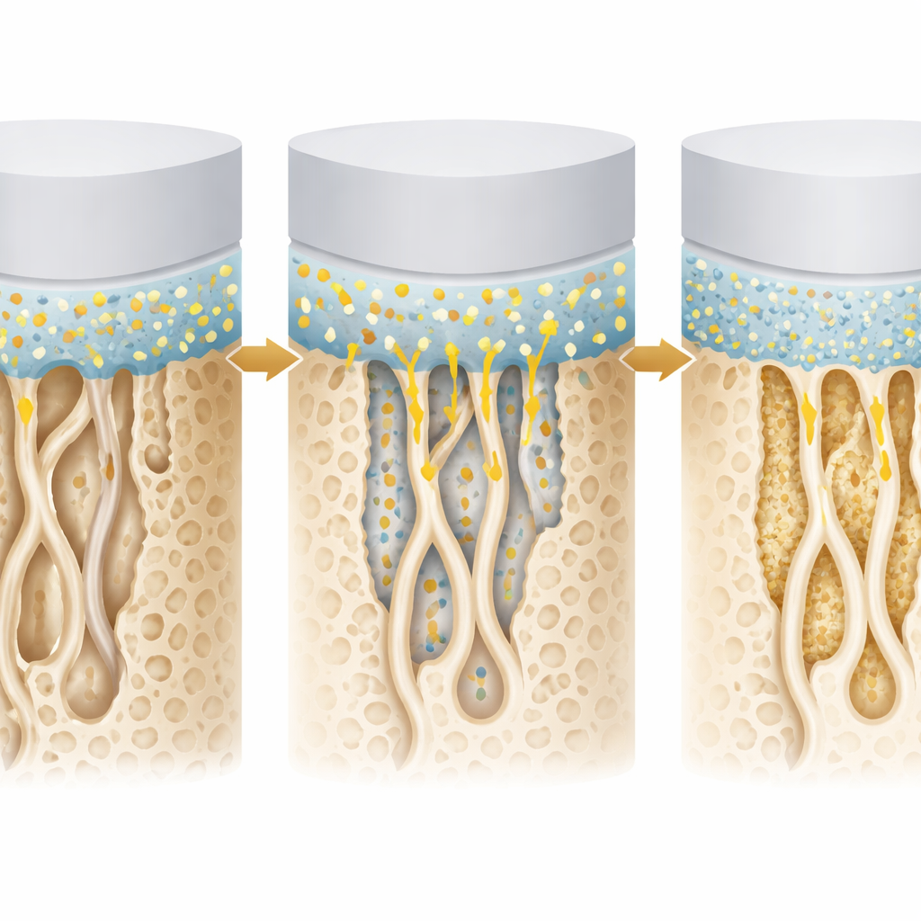
Today’s tooth-colored fillings rely on adhesives that bond plastic-based composite to dentin. This bond forms a thin contact zone where tooth collagen fibers and liquid resin weave together, often called the hybrid layer. Over time, water, chewing forces, and natural enzymes can break down this delicate region, weakening the bond and allowing bacteria or fluids to seep in. Finding ways to protect or even rebuild this layer could mean fewer failed fillings and better preservation of healthy tooth structure.
A mineral twist on a universal adhesive
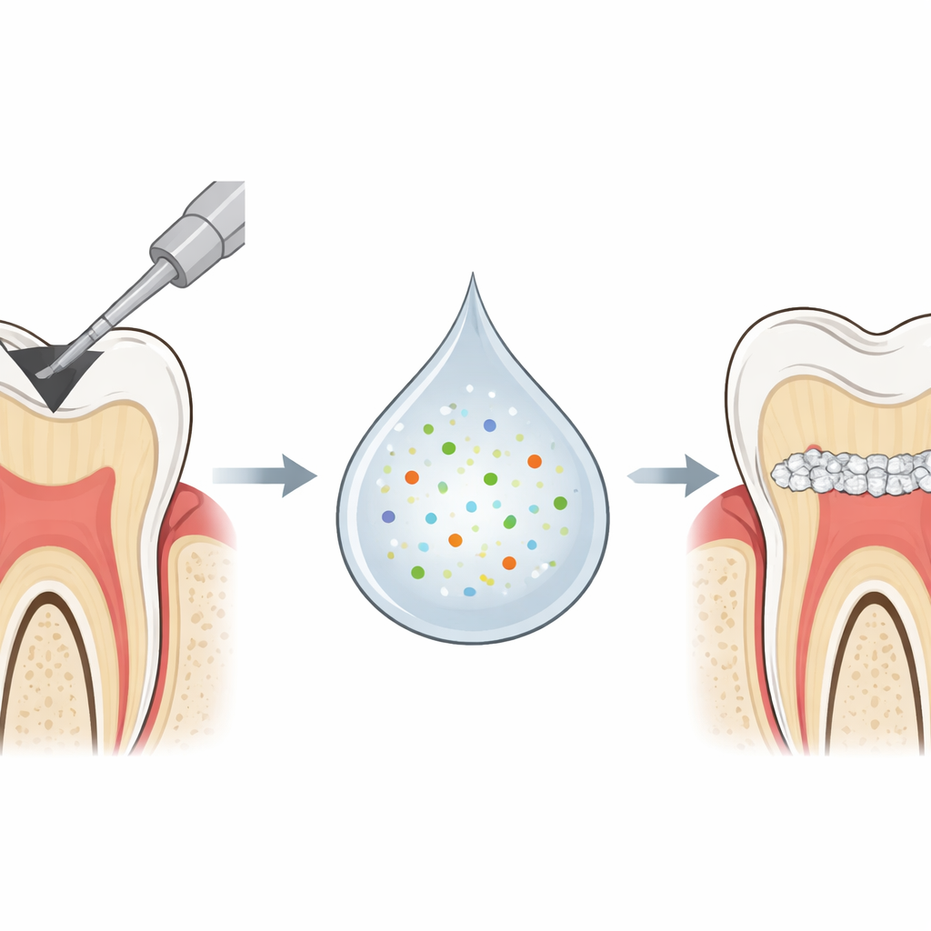
In this research, dentists started with a widely used “universal” adhesive—a single liquid that can be applied in a simple self-etch manner to dentin. They then mixed in a very low amount (0.5% by weight) of tricalcium silicate powder, a bioactive mineral used in other dental repair materials. This mineral is known to react with water, releasing calcium and creating new mineral deposits. Forty extracted human molars were prepared with flat dentin surfaces and restored with composite fillings using either the standard adhesive or the mineral‑enhanced version. Some teeth were tested after one day, while others were stored in water for six months to simulate aging.
Putting the bond to the test
To measure how well the fillings held up, the researchers cut each restored tooth into tiny beams and gently pulled them apart until the bond failed, recording the microtensile bond strength. They also examined the tooth–filling interface under a scanning electron microscope to see how well the adhesive had penetrated and how the layers looked over time. Immediately after placement, both the standard and the tricalcium silicate–modified adhesives showed similar bond strengths. However, after six months in water, the story changed: the bond strength of the standard adhesive dropped sharply, while the mineral‑enhanced adhesive not only maintained its strength but showed the highest values among all groups.
What happens at the contact zone
Microscope images revealed thin bonding layers and short resin extensions into the dentin for both adhesives, features that are common with self‑etch systems. The mineral‑modified adhesive sometimes showed small gaps along the interface, yet this did not translate into weaker bonds. Most failures occurred right at the adhesive layer, especially in the standard adhesive after aging, which matched its lower strength values. In contrast, the tricalcium silicate groups had fewer purely adhesive failures, suggesting a more resilient contact zone between filling and tooth. The authors suggest that, as the mineral particles hydrate, they may release calcium and form new mineral deposits, helping to stabilize the collagen and resist breakdown in a wet environment.
What this could mean for your next filling
In simple terms, adding a small amount of tricalcium silicate to a common dental adhesive helped the bond to dentin stay strong over six months of water storage, while the regular adhesive weakened. Although these tests were done in the lab and not in patients’ mouths, the results hint that “smart” adhesives containing bioactive minerals could lead to longer‑lasting white fillings by supporting natural mineral repair at the tooth–filling seam. If future studies in real-world conditions confirm these findings, dentists may one day use such enhanced adhesives to keep restored teeth stronger and healthier for longer.
Citation: Zayed, T., Elkholany, N., Motawea, A. et al. Effect of incorporation of tricalcium silicate to a universal adhesive on microtensile bond strength to dentin and micromorphological patterns of tooth/restoration interface. Sci Rep 16, 12025 (2026). https://doi.org/10.1038/s41598-026-44781-1
Keywords: dental adhesive, tooth fillings, dentin bond strength, bioactive materials, tricalcium silicate