Clear Sky Science · en
Analysis of the kinematic features of pelvic and lower limb motion in patients with hip osteoarthritis and acetabular dysplasia
Why hip motion matters in everyday walking
Many young adults develop painful hip arthritis long before old age because the socket of the hip joint is too shallow, a condition called acetabular dysplasia. This shallow socket makes the hip less stable and pushes the body to find workarounds every time a person takes a step. The study in this article explores how such people walk in three dimensions, from pelvis to ankle, to uncover subtle motion patterns that could guide surgery and rehabilitation and help them move with less strain and more comfort in daily life.
Looking closely at hips that wear out early
The researchers focused on 25 young adults with hip osteoarthritis caused by acetabular dysplasia and compared them with 25 healthy people of similar age, height, and walking speed. Instead of relying only on simple measurements like the biggest bend of a joint, they used a motion capture system similar to those used in film and video games. Reflective markers placed on the body and force plates in the floor allowed them to track how the pelvis, hips, knees, and ankles moved in space during walking. They then analyzed these continuous movement traces through the entire step, rather than just at a few selected moments.
How the pelvis and hip change their role
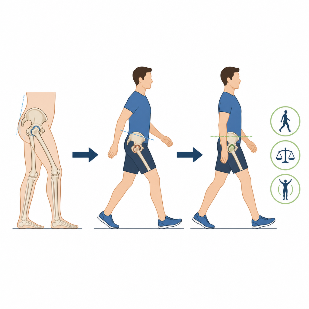
Compared with the healthy group, people with dysplasia walked with their pelvis tipped more forward and with less ability to tilt backward. As they stepped, the side of the pelvis that was not on the ground dropped more, showing a pattern often seen in people with weak or overloaded hip muscles. Their hips bent more but straightened less, meaning they avoided moving the leg far behind the body. This combination suggests that they may be protecting the front of the hip from painful forces by limiting backward swing, while using extra bending at the hip to keep their stride and speed close to normal.
Surprising help from the ankle and foot
The study also found that the ankle and foot played a larger supportive role than expected. People with dysplasia showed less downward pointing of the foot during push off and more outward rolling of the ankle through much of the step. This outward roll is related to foot pronation, flat feet, and toe deformities, which are more common in these patients. These changes likely help redirect forces and keep balance when the hip joint is not well covered by the socket. Interestingly, the knee moved quite similarly in both groups, suggesting that the body leans more on the pelvis and ankle for compensation than on the knee itself.
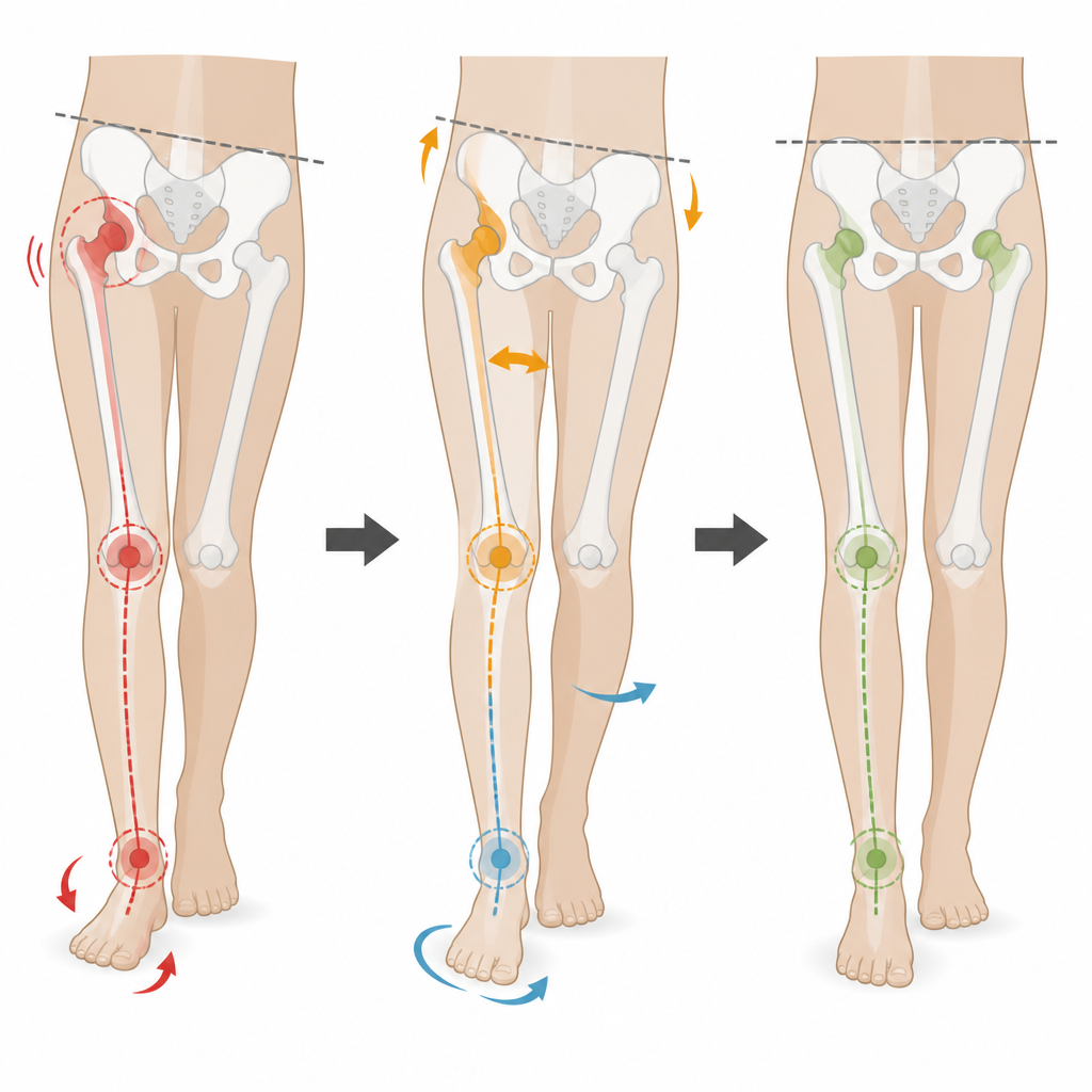
Connecting hip problems to whole leg motion
By combining traditional peak measurements with time based statistical mapping, the authors showed when during the step these altered movements occur and how long they last. They link the shallow socket shape and rotated thigh bone seen in dysplasia to a chain of effects: forward tilt of the pelvis, reduced hip extension, inward twist of the leg, and greater ankle eversion. This chain supports the idea of a close “hip ankle relationship,” where changes at the hip and ankle are tightly coordinated as the body tries to stabilize an unstable joint.
What this means for care and recovery
For a layperson, the take home message is that a problem in the hip does not stay in the hip. A shallow socket changes how the pelvis tips, how far the leg swings, and how the ankle and foot roll with each step. These shifts are not random but appear to be the body’s way of protecting a vulnerable joint. Understanding these patterns in detail can help surgeons better plan how to reshape or replace the hip and guide therapists to include pelvis and ankle training, not just hip exercises, in rehabilitation. In the long run, such tailored care may improve comfort and function for young people living with this form of early hip arthritis.
Citation: Ueki, S., Shoji, T., Iwamoto, Y. et al. Analysis of the kinematic features of pelvic and lower limb motion in patients with hip osteoarthritis and acetabular dysplasia. Sci Rep 16, 15689 (2026). https://doi.org/10.1038/s41598-026-44774-0
Keywords: hip osteoarthritis, acetabular dysplasia, gait analysis, pelvis and ankle motion, compensatory walking