Clear Sky Science · en
Laboratory-based cellular-level correlative visible-light and X-ray microscopy for 3D evaluation of mouse kidney biopsy
Why looking inside tiny kidney samples matters
A kidney biopsy is a sliver of tissue that can reveal why someone’s kidneys are failing, but today doctors mostly see it as a stack of flat slices under a light microscope. This study explores a way to look at that same sample in three dimensions, without cutting it apart, by pairing X-ray microscopy with familiar light microscopy. The goal is a fuller picture of kidney damage, especially in the tiny filters called glomeruli, using tools that could fit into ordinary lab workflows.
Limits of current ways of seeing
Traditional kidney pathology relies on very thin, stained sections viewed with light or electron microscopes. These methods can show individual cells and fine structures but require cutting the biopsy into many slices. That process is time consuming, destroys the original shape of the tissue, and usually covers only a small portion of the sample. Even when stacks of slices are used to reconstruct a three-dimensional view, the spacing between them can blur details in depth. Newer light-sheet methods can scan intact tissue in 3D but only show parts of the sample that glow, leaving much of the structure invisible.
A new paired imaging approach
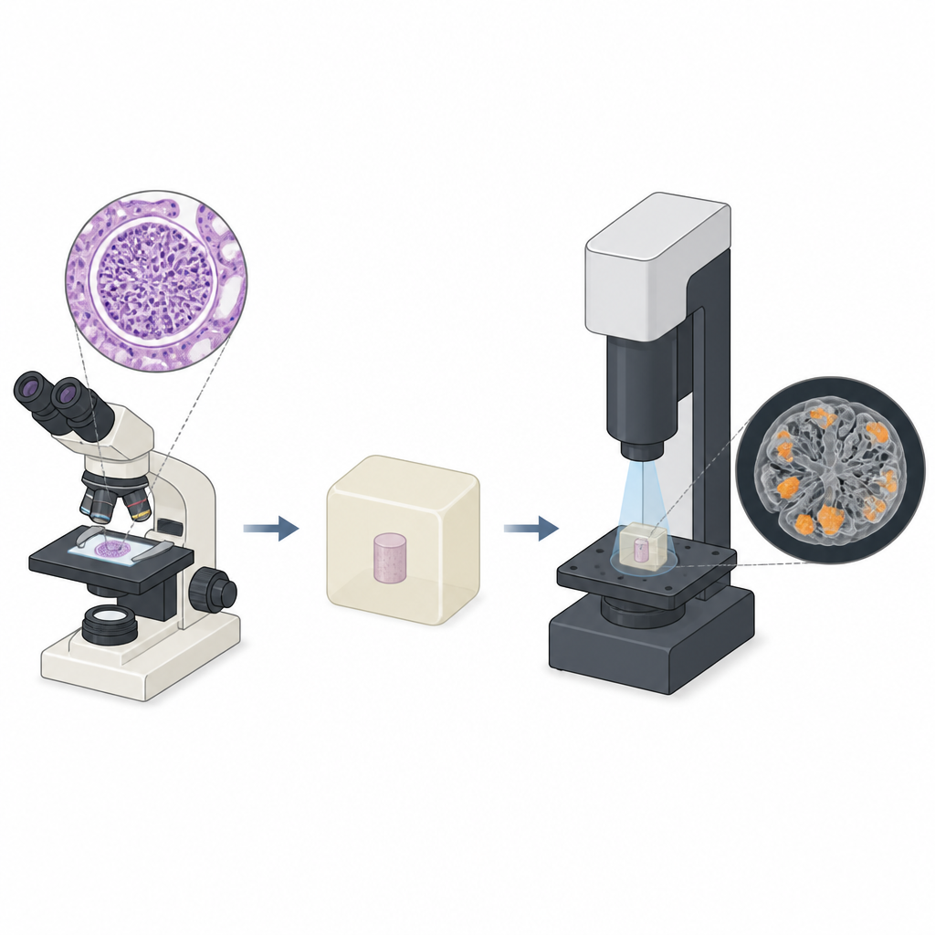
The researchers built on earlier work showing that standard wax used in tissue processing can help X-rays distinguish cells inside an uncut biopsy. In this study they introduced what they call laboratory-based cellular-level correlative light and X-ray microscopy. First, they scanned a wax-embedded mouse kidney biopsy with an X-ray microscope to create a 3D image at cell-level resolution. They then processed the same piece of tissue for routine staining and light microscopy at matched locations. By carefully aligning the two data sets, they could compare features cell by cell and use the strengths of each method together.
Sharpening the X-ray view
To make the X-ray pictures clear enough to see individual cell nuclei, the team had to solve several technical problems. Long scans made the sample shift slightly as the room temperature changed, creating streaky artifacts. They attached a tiny hard particle to the sample as a position marker and used its motion to correct for drift in the raw X-ray images. They also applied a mathematical step called phase retrieval to boost contrast between tissue and wax. Together, these steps greatly improved image sharpness and contrast, making it possible to spot dense spots that corresponded to cell nuclei in different parts of the nephron, such as tubules and glomeruli.
Matching cells between light and X-ray images
With clearer X-ray data, the scientists aligned the 3D X-ray slices with 2D stained light microscope images by using cell nuclei as landmarks. When they compared matched regions, they found that bright, dense spots in the X-ray images lined up with nuclei seen in stained sections. This allowed them to confidently identify different cell types and regions, including normal supporting areas inside glomeruli and clusters of extra cells known as hypercellularity. They also noticed that the cut and stained sections showed more local distortion than the intact X-ray volumes, likely due to the physical handling of the tissue during standard preparation.
Revealing hidden three-dimensional lesions
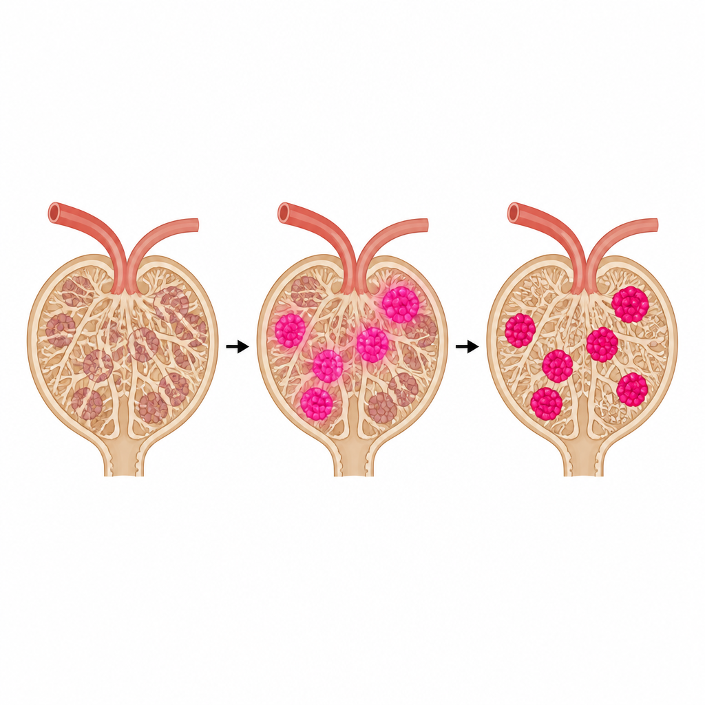
The team focused on a disease-model mouse in which the remaining kidney develops injury in its glomeruli. Using the paired images, they manually outlined one glomerulus and its dense internal regions in the X-ray data, guided by the stained sections. They identified three distinct hypercellular lesions, each with a different 3D shape: a simple bump off a normal branch, a fused cluster of bumps, and a cramp-like bridge between branches. By counting nuclei on light images and measuring volumes in the X-ray data, they estimated that each mesangial cell in these clusters occupied roughly 100 cubic micrometers. While based on a single glomerulus, these measurements match general expectations from pathology and show that such volumetric information can be extracted from intact biopsies.
What this could mean for kidney diagnosis
This work shows that an ordinary wax-embedded kidney biopsy can be scanned by a lab-based X-ray microscope to yield a detailed 3D map of its tiny filters, then reused for standard staining and light microscopy. Used together, the two methods make it possible to pinpoint and measure cell-rich lesions that may signal disease, without destroying the overall structure of the sample. Although the current process is slow and relies on manual steps, future automation and better software could turn this into a practical tool, giving clinicians a more complete 3D view of kidney damage from the same small piece of tissue they already collect.
Citation: Kunishima, N., Hirose, R., Takeda, Y. et al. Laboratory-based cellular-level correlative visible-light and X-ray microscopy for 3D evaluation of mouse kidney biopsy. Sci Rep 16, 15634 (2026). https://doi.org/10.1038/s41598-026-44720-0
Keywords: kidney biopsy, X-ray microscopy, 3D imaging, glomerulus, mesangial cells