Clear Sky Science · en
Comparative histological and histochemical analysis of the minor salivary glands in porcine species
Why tiny mouth glands in pigs matter
Every time you chew, speak, or swallow, a hidden network of tiny glands quietly coats your mouth with saliva. This thin protective film keeps food sliding smoothly, shields your teeth, and helps control germs. In humans, when these glands fail—after radiation therapy, autoimmune disease, or aging—life can become painfully dry. The study described here takes a close look at the small, often overlooked salivary glands in pigs and shows why they are such a powerful stand-in for understanding and treating human dry mouth and related oral diseases. 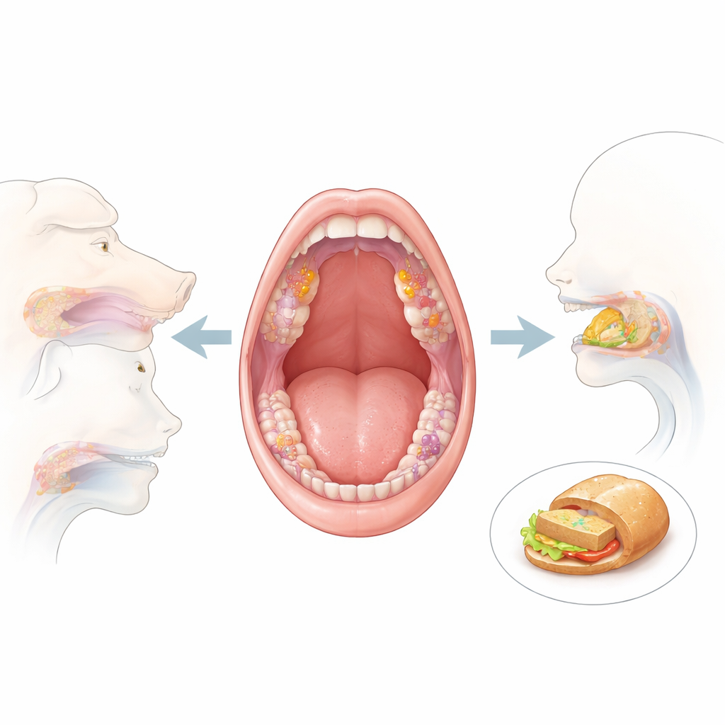
Hidden moisture factories in the mouth
Saliva does not come from just the big, well-known glands near your jaw. Scattered through the lips, cheeks, tongue, and soft palate are hundreds of minor salivary glands, each only a fraction of a millimeter across. They continuously drip saliva directly onto the surface of the mouth, forming a protective layer even when you are not eating. In this work, researchers collected heads from ten healthy adult pigs at a slaughterhouse and systematically sampled 18 well-defined spots across the cheeks, lips, tongue, and palate. Using thin tissue slices stained with a range of dyes, they mapped where these tiny glands sit, what they look like, and what kind of saliva they produce.
Different glands for different jobs
The team found that these minor glands are not identical copy-and-paste units. Instead, each region in the pig’s mouth has its own pattern of glands tuned to local needs. In the cheeks, two clearly separated rows of glands run along the inside: a dorsal (upper) row that produces thick, mucous-rich saliva, and a ventral (lower) row that makes a much thinner, watery secretion. Elsewhere—the tongue, soft palate, and lips—glands are mostly mucous-secreting, with occasional patches of watery units or mixed clusters where watery cells cap a mucous core. Under the large taste buds at the back of the tongue, special “gustatory” glands produce a non-mucous, enzyme-rich fluid thought to help flush and reset taste surfaces between bites.
What the dyes reveal about their chemistry
To understand what these glands actually secrete, the scientists turned to classic histochemical stains, each binding to specific chemical groups. Mucous-producing cells in most regions lit up strongly with dyes that mark both neutral and acidic sugar-rich molecules—glycogen, sialomucins, and sulfated mucins. These sticky, highly hydrated compounds create a smooth, slippery, and protective film over the mouth’s lining, helping food form a bolus, cushioning the tissue against abrasion, and trapping microbes. In sharp contrast, the watery serous cells and their ducts were consistently negative for these mucin-focused stains, but showed structural traits of protein factories, such as dark, grainy cytoplasm. This pattern supports the idea that they specialize in enzyme and protein secretion rather than thick mucus. 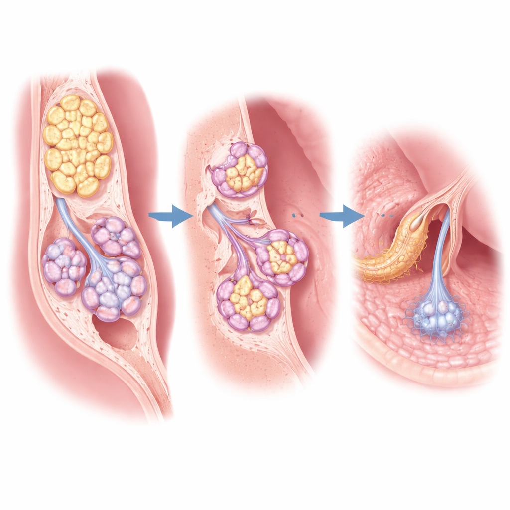
Shape and size tuned to flow
Beyond chemistry, the researchers measured the diameters of gland units and their ducts across regions and types. Mucous acini—the rounded clusters of mucous cells—were significantly larger and had wider central spaces than their serous counterparts, consistent with storing and releasing viscous material. Ducts draining mucous glands in the tongue, for example, were markedly wider than those serving serous glands in the cheek, reflecting higher volumes and different flow properties. A careful statistical analysis, which treated each animal as its own cluster of measurements, confirmed that these size differences across sites were highly significant. The overall layout was similar everywhere—small collecting ducts feeding into larger channels that eventually open onto the mouth surface—but the dimensions shift depending on how much fluid and what type of fluid needs to be moved.
Why this matters for human health
Although this work focuses on pigs, its implications reach directly into human medicine. Pigs share many structural and functional features of the mouth with people, making them an excellent “testbed” for understanding how saliva is produced and how it can be restored when it fails. By providing a detailed baseline map of where each type of minor gland is located, what it secretes, and how its duct system is built, this study offers a reference for surgeons, pathologists, and tissue engineers. It supports the use of pig tissue as a scaffold for growing replacement glands and as a realistic model for testing new treatments for conditions like radiation-induced dry mouth or autoimmune damage. In simple terms, the researchers show that the pig’s tiny mouth glands closely mirror our own—and that understanding them in detail may help keep human mouths moist, protected, and comfortable throughout life.
Citation: Rao, P., Singh, A., Kumar, P. et al. Comparative histological and histochemical analysis of the minor salivary glands in porcine species. Sci Rep 16, 14347 (2026). https://doi.org/10.1038/s41598-026-44696-x
Keywords: salivary glands, pig model, oral health, dry mouth, mucus secretion