Clear Sky Science · en
Calcaneal spur detection from lateral foot radiographs using deep feature engineering
Why sore heels and smart computers matter
Heel pain is one of the most common reasons people visit orthopedic clinics, often making every step uncomfortable. A frequent culprit is a tiny bony outgrowth on the heel called a calcaneal spur, usually seen on standard side-view foot X-rays. Reading these images can be surprisingly tricky, especially in early disease, and not every hospital has expert radiologists on hand. This study introduces a streamlined computer method that can reliably spot these spurs on ordinary X-rays, even when only a modest number of images are available, potentially offering faster, more consistent help for doctors and patients.
What heel spurs are and why they are hard to see
Calcaneal spurs form where the strong band of tissue under the foot, the plantar fascia, attaches to the heel bone. Age, excess weight, long periods of standing, and repeated small injuries all raise the chance of developing them. Doctors usually begin their evaluation with a simple lateral foot X-ray because it is quick, inexpensive, and widely available. The problem is that early spurs can look very similar to normal heel shapes, and image quality or slight changes in foot position can also create spur-like shadows. Different radiologists may interpret the same X-ray differently, which is where computer-aided systems can offer a second opinion.
A new way to teach computers from few examples
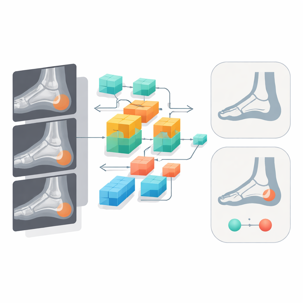
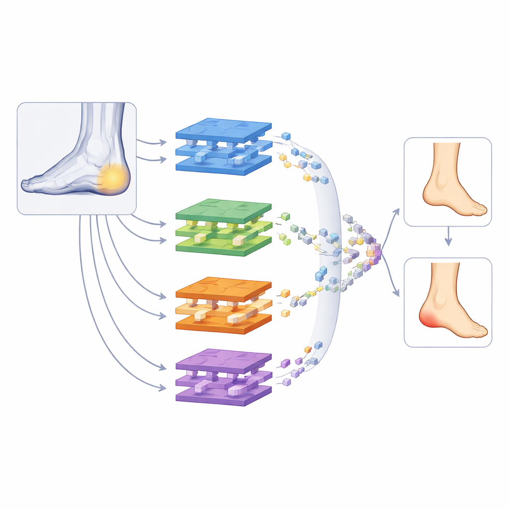
Modern artificial intelligence systems known as deep learning have transformed medical image analysis, but they typically need thousands of labeled images and powerful graphics processors to work well. In real clinics, especially for specific conditions like heel spurs, only a few hundred well-curated images may be available and computing resources can be limited. The authors tackle this challenge with what they call deep feature engineering: instead of training one very large model end-to-end, they reuse knowledge from 19 existing image-recognition networks and then carefully select and combine what each has learned. This strategy aims to capture rich visual cues while keeping the overall system light enough for everyday use.
How the multi-step system works
The team collected 775 de-identified lateral foot X-rays from a single hospital, of which 316 showed no spur and 459 showed a calcaneal spur, as agreed upon by two experienced clinicians. Each image was resized but otherwise left intact so that the whole foot, not just a cropped region, was analyzed. First, every image was passed through 19 different pre-trained neural networks, each originally trained on a large general-purpose image database. From each network, the researchers extracted a numerical fingerprint, or feature set, summarizing how that network “sees” the heel region. Next, they used an iterative selection method to trim each large fingerprint down to a smaller subset of the most informative features, discarding redundant or noisy measurements that did not help distinguish spur from no spur.
Combining many small votes into one strong decision
Once the leaner feature sets were ready, they were fed to a special variation of a simple, human-intuitive classifier called k-nearest neighbors, which judges a new case by looking at the most similar past examples. The authors expanded this idea into an ensemble: they tried many distance settings and neighbor counts and then used repeated majority voting to fuse the numerous candidate decisions. A final greedy step selected whichever combination performed best during cross-validation tests. Overall, the framework reached an accuracy of 93.42 percent, with particularly high sensitivity for cases that actually had spurs. In other words, it was more likely to miss a normal heel than to overlook a spur, which can be a safer balance in a screening context.
What the results mean beyond heel pain
To ensure the system was looking at the right place, the researchers visualized which regions the networks focused on and found strong attention around the underside of the heel, where spurs are expected to form. They also showed that carefully combining several networks beat any single one on its own, and that similar gains appeared when the framework was applied to a separate, more complex cell-image dataset. Because the method runs on a standard computer without a graphics card and uses only a handful of networks at deployment, it could realistically be added to hospital workflows as a background assistant. For patients, this does not replace expert judgment, but it could mean more consistent readings of common X-rays, quicker reassurance when no spur is present, and better support for doctors who must decide how best to manage stubborn heel pain.
Citation: Demir, S., Can, B., Goktas, O.F. et al. Calcaneal spur detection from lateral foot radiographs using deep feature engineering. Sci Rep 16, 14125 (2026). https://doi.org/10.1038/s41598-026-44671-6
Keywords: heel pain, X-ray imaging, medical AI, bone spur, computer-aided diagnosis