Clear Sky Science · en
Intraindividual comparison of photon-counting detector CT and third-generation dual-source energy-integrating detector CT in multiphasic contrast-enhanced abdominal CT
Clearer Scans for a Common Test
CT scans of the abdomen are a routine tool for finding and tracking cancers, liver disease, and other serious conditions. This study asks a simple but important question: can a new type of CT scanner make these images clearer without exposing patients to more radiation? By directly comparing two scanner designs in the same group of patients, the researchers show that a newer "photon-counting" CT system can sharpen key details inside the abdomen, potentially helping doctors spot small or subtle problems earlier.
Two Ways to Catch the Same X-rays
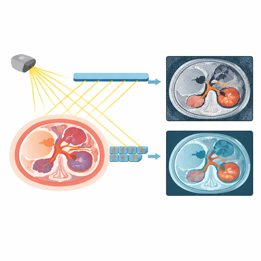
Most hospitals today use CT scanners that add up the total energy of X-rays passing through the body. The new scanners tested here take a different approach: they count individual X-ray particles and record their energy one by one. This difference might sound technical, but it matters because it can boost how strongly iodine contrast dye shows up and reduce unwanted background noise. The team focused on multiphasic abdominal scans, which are taken at two time points after injection of contrast dye—an arterial phase and a portal venous phase—because these are critical moments for seeing how blood flows through organs such as the liver and pancreas.
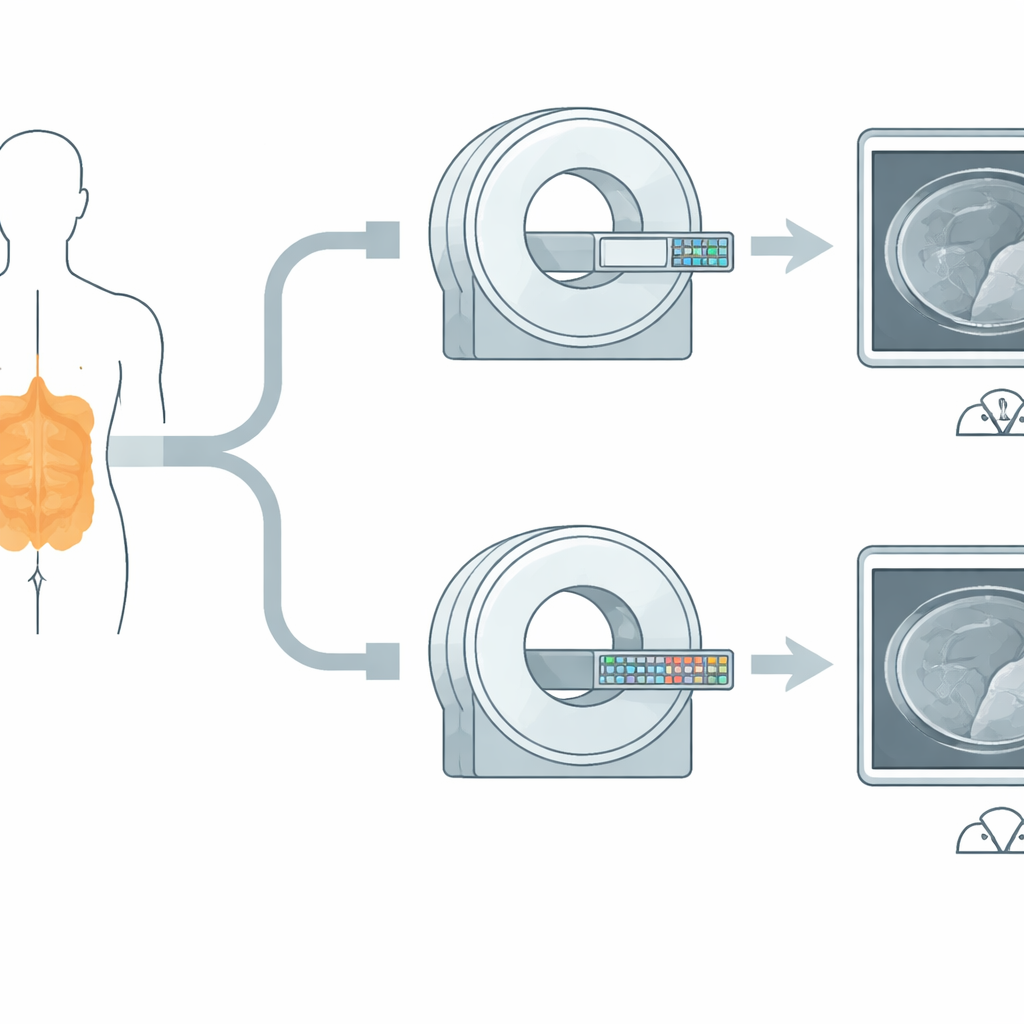
A Head-to-Head Test in Real Patients
The researchers looked at 88 patients who, for clinical reasons, had undergone contrast-enhanced abdominal CT on both a state-of-the-art conventional scanner and a new photon-counting scanner within about a year. They kept key factors as similar as possible: both systems used the same X-ray setting, and the photon-counting scans were tuned so that the radiation dose closely matched that of the conventional exams. From the photon-counting data, they generated special images that mimic X-rays of different energies, called virtual monoenergetic images, at several energy levels. This allowed them to see which settings gave the best balance of organ visibility and image smoothness.
Sharper Contrast Where It Counts
Using precise measurements in the aorta, portal vein, liver, pancreas, and back muscles, the team found that photon-counting images at lower energies (especially 60–70 keV) made iodine-filled blood vessels and organs appear brighter relative to their surroundings. At 70 keV, these images showed 13–50 percent higher contrast-to-noise ratios than conventional CT, depending on the organ and phase, meaning important structures stood out more clearly against background noise. Image graininess decreased as energy increased, but even at 70 keV the photon-counting images were less noisy than the standard scans. Very low energies (50 keV) produced the strongest contrast but also the most graininess, making them useful for selected tasks but not ideal for routine viewing.
Radiologists’ View from the Reading Room
Two experienced abdominal radiologists rated the images without knowing which scanner produced them. They judged vessel contrast, organ contrast, image noise, and overall quality on a five-point scale. Their scores agreed well with each other and with the numerical results: photon-counting images at 60 and 70 keV received the highest overall ratings, clearly beating the conventional CT images at the same dose. Images at 50 keV, 80 keV, and 90 keV were judged less favorable overall, either because of excess graininess (at low energy) or loss of contrast (at high energy). The study thus identified a sweet spot where the new scanner’s strengths translate into images that are both sharp and comfortable to read.
What This Means for Patients
For patients, the key message is that the photon-counting CT system can produce clearer abdominal images without increasing radiation exposure, and sometimes with a slightly lower dose. Stronger contrast in blood vessels and organ tissue may help doctors detect small liver or pancreatic tumors and see fine blood vessel details that guide surgery or other treatments. While the work did not directly measure how often cancers or other diseases were found, it lays the groundwork for future studies that will link these sharper images to real-world diagnostic gains. If those benefits are confirmed, photon-counting CT could become an important upgrade to one of medicine’s most widely used imaging tests.
Citation: Sofue, K., Nishiuchi, K., Ishikawa, K. et al. Intraindividual comparison of photon-counting detector CT and third-generation dual-source energy-integrating detector CT in multiphasic contrast-enhanced abdominal CT. Sci Rep 16, 10975 (2026). https://doi.org/10.1038/s41598-026-44618-x
Keywords: photon-counting CT, abdominal imaging, contrast-enhanced CT, liver and pancreas lesions, medical imaging technology