Clear Sky Science · en
Automated detection and segmentation of Weiss ring in fundus photography images using deep learning
Why tiny rings in the eye matter
As people age, many notice new “floaters” drifting across their vision. Most are harmless, but some can hint at changes that occasionally lead to retinal tears and even blindness. One of the clearest signs of such change is a small, ring-shaped speck inside the eye called a Weiss ring. In everyday eye photos, however, this ring can be faint and easy to miss—even for experts, and especially for computers. This study describes a new artificial intelligence (AI) system that learns to spot and outline Weiss rings in standard retinal photographs, potentially turning routine images into a richer source of clinical and research insight.
A subtle clue inside routine eye photos
The inside of the eye is filled with a clear gel that slowly separates from the retina as we get older, a process known as posterior vitreous detachment. When this separation is complete, it can leave behind a floating loop of tissue in front of the optic nerve head: the Weiss ring. Its presence signals that the separation has finished, and in some patients with sudden flashes or floaters, it can mark a time window when the retina is more vulnerable to small tears. Yet Weiss rings are not always visible, can fall outside the camera’s field of view, and often look like vague shadows. Traditional AI systems trained to find common retinal diseases have mostly ignored these structures or misread them as artifacts, leaving an important piece of information unused.
Teaching computers to find the ring
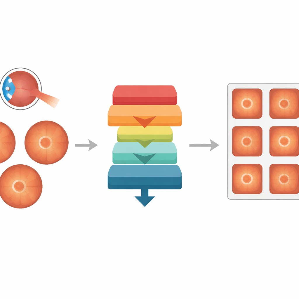
The researchers assembled 582 color eye photographs from several public databases and asked three eye specialists to label which images contained a Weiss ring and to carefully trace its outline. This produced 194 images with rings and 388 without. They then built a two-part AI system. One part, called a segmentation network, learned to draw a pixel-by-pixel map of where the ring sits in each image. The other part, a classification network, learned to decide whether a ring is present in the picture at all. A second classifier examined only the segmentation mask, judging whether the marked area truly looked like a ring. Finally, a simple statistical model combined the two classification scores into a single, final prediction.
How well the system performed
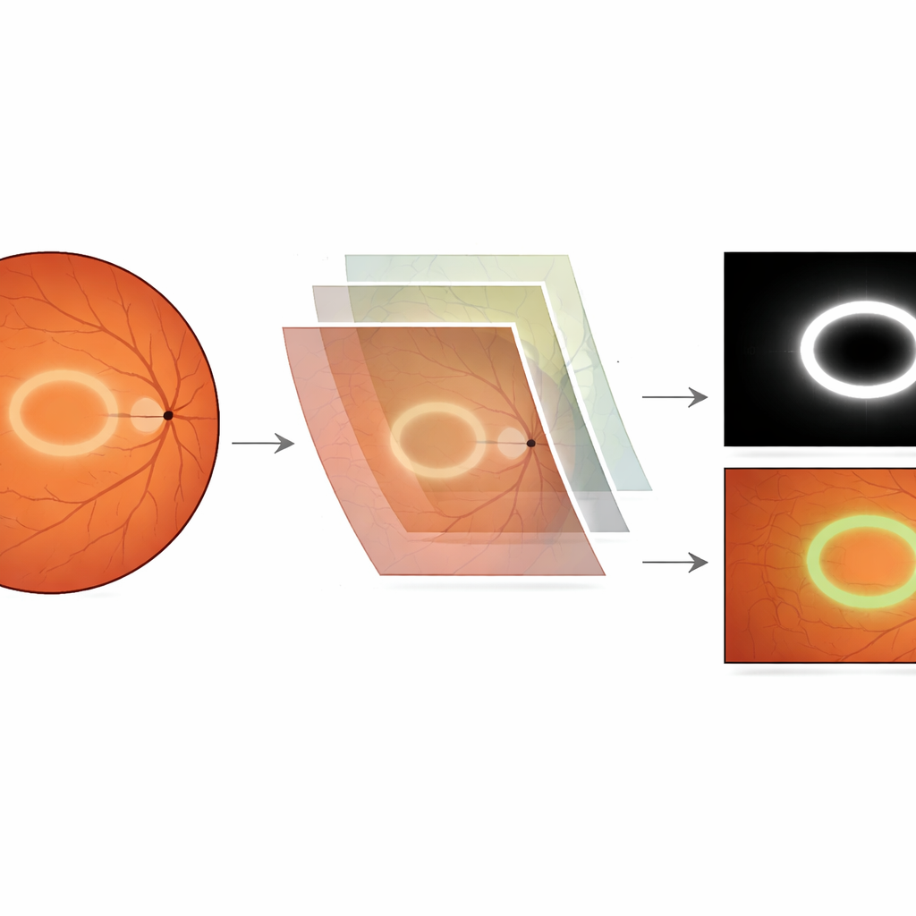
On a held-out test set that the AI never saw during training, the ring-outlining network produced maps that overlapped reasonably with the experts’ drawings, especially in clearer cases. It was particularly sensitive, catching most true rings, though it sometimes marked extra regions where reflections or background texture mimicked a ring. When the segmentation output was fused with the image-level classifier, overall detection improved. The combined system correctly distinguished ring from no-ring images with a high area-under-the-curve value (about 0.90) and solid accuracy, while also showing good ability to rule out images without a ring. Visual heatmaps confirmed that the model mainly focused on the area around the optic nerve where Weiss rings typically appear, adding confidence that it was using clinically meaningful cues rather than spurious patterns.
What this could mean for care and research
The authors stress that this tool is not a replacement for a full eye exam or advanced scans when someone reports sudden flashes or floaters. Standard photographs cover only part of the retina, and many people with vitreous detachment never show a visible ring. Instead, the system is presented as an assistant: it can flag images for closer review in large screening programs, help trainees learn to recognize subtle rings, and enable researchers to sift through thousands of archived photos to study how often rings occur and how they relate to factors like age, surgery, or other eye diseases. Because the study used a modest number of publicly sourced images, larger, real-world trials—ideally comparing results with wide-angle imaging or three-dimensional scans—will be needed before the approach can be trusted in everyday clinic decisions.
A small ring with big data potential
In plain terms, this work shows that a computer can be trained not only to say “yes or no” about a tiny floating ring in the eye, but also to sketch where it is. While the system is still a proof of concept and not a diagnostic authority, it opens the door to turning an often-overlooked feature into a searchable signal across huge image collections. Used carefully and alongside proper examination, such AI could help doctors and researchers pay more attention to subtle floaters that tell a story about the aging eye.
Citation: Kim, H., Ryu, S.Y., Yoo, T.K. et al. Automated detection and segmentation of Weiss ring in fundus photography images using deep learning. Sci Rep 16, 13787 (2026). https://doi.org/10.1038/s41598-026-44593-3
Keywords: Weiss ring, retinal imaging, deep learning, vitreous detachment, medical AI