Clear Sky Science · en
Isobaric quantitative proteomics reveals altered extracellular matrix, cytoskeletal, and degradation pathways in glaucomatous trabecular meshwork cells
Why this matters for vision
Glaucoma is a leading cause of irreversible blindness, largely because pressure inside the eye slowly damages the optic nerve. That pressure depends on how easily a clear fluid can drain through a tiny sieve-like tissue called the trabecular meshwork. This study digs deep into the proteins inside cells from this drainage tissue in people with and without glaucoma, revealing how their internal machinery shifts in disease and pointing to fresh ideas for preventing vision loss. 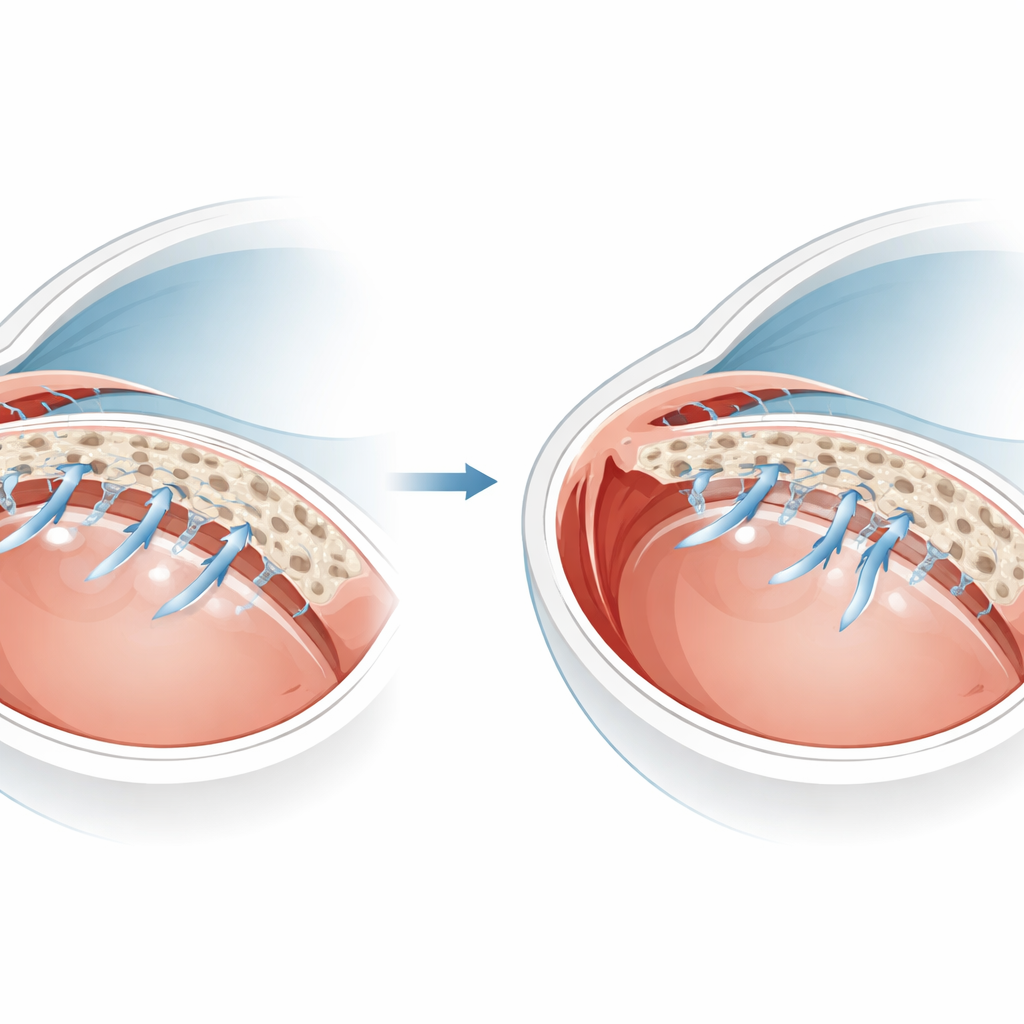
The eye’s clogged drain
In healthy eyes, fluid produced inside the eye flows forward and escapes through the trabecular meshwork, a layered filter that lines the edge of the cornea. Specialized cells within this tissue constantly remodel their surroundings so the fluid can pass at just the right rate to keep pressure stable. In glaucoma, this balancing act fails: the tissue accumulates a tangled, stiff extracellular matrix, the cells become fewer and more rigid, and pressure creeps upward. Because human eye tissue is scarce and tiny, it has been hard to measure all the proteins involved. The authors turned to cultured cells grown from donor eyes with and without glaucoma, which preserve many of the features of the original tissue, and used a highly sensitive mass‑spectrometry method to compare thousands of proteins at once.
Profiling thousands of tiny parts
Using a technique called tandem mass tag proteomics, the team labeled protein fragments from five glaucoma-derived cell strains and five matched non‑glaucoma strains. This allowed them to run all samples together in a single, carefully controlled experiment and then read out how abundant each protein was in every sample. They detected more than 5,500 proteins and found 248 that consistently differed between glaucoma and control cells: 206 were increased and 42 decreased in glaucoma cells. Computer analyses grouped these proteins into major biological themes, highlighting changes in the material surrounding the cells, the internal scaffolding that shapes them, how they break down worn‑out components, and proteins associated with the cell nucleus and nucleolus.
Stiffer surroundings and tenser cells
One major shift involved the extracellular matrix—the mesh of collagen and other molecules that forms the physical filter for fluid. Glaucoma cells produced more of some matrix proteins, such as a collagen fragment called arresten, and less of others like decorin, which normally helps organize collagen fibers. They also altered adhesion molecules and components of Wnt signaling, a pathway that helps cells sense and respond to their environment. Inside the cells, many proteins tied to the actin cytoskeleton—the internal cables that control shape and contractility—were elevated. These included ROCK2, a key enzyme already targeted by a newer class of glaucoma drugs, as well as moesin, tropomyosin‑2, cofilins, and vimentin. Together, these changes support the idea that glaucoma cells are more contractile and mechanically stressed, further tightening the eye’s drainage system. 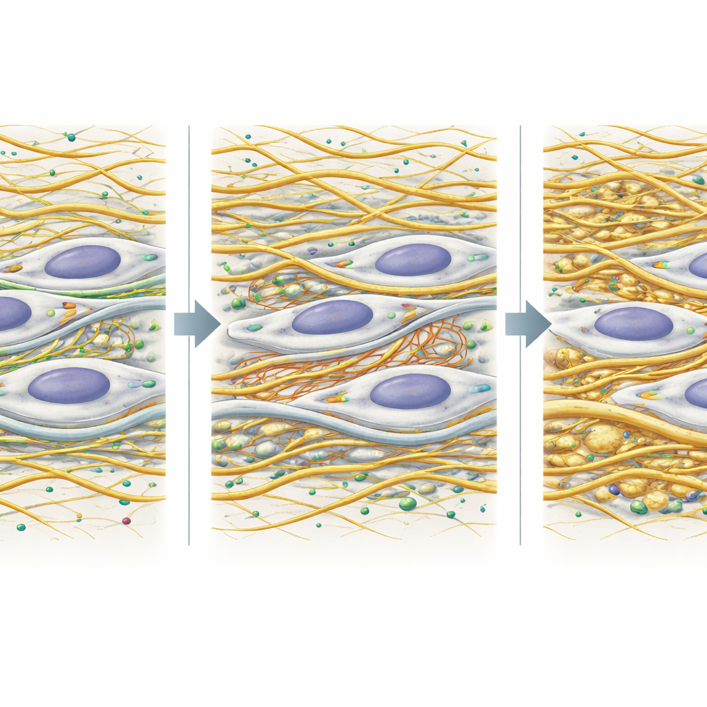
Strained waste disposal and altered nuclei
The study also uncovered signs that the cells’ waste‑processing machinery is out of balance. Components of both the ubiquitin–proteasome system, which tags and shreds damaged proteins, and the autophagy–lysosome pathway, which digests larger cellular debris, were shifted up or down in glaucoma cells. For example, enzymes that add or remove “trash tags” on proteins, and a key lysosome membrane protein, were all increased, hinting at a stressed but imperfect cleanup system. At the same time, several nuclear proteins rose sharply, including lamin A/C, which helps maintain the shape and stiffness of the nucleus, and SNX7, which the authors found in enlarged nucleoli—the cell’s ribosome factories—of glaucoma cells. These enlarged nucleoli and nuclear changes align with broader ideas about cellular aging and stress in glaucoma.
What this means for future treatments
By mapping how dozens of protein networks shift in glaucoma drainage cells, this work confirms that the disease is not driven by a single culprit but by coordinated changes in the tissue scaffold, cell mechanics, and cellular housekeeping. The findings reinforce current drug strategies that relax the cytoskeleton and open the eye’s drain, while pointing to new targets in matrix organization, waste‑disposal pathways, and nuclear structure. For patients, the takeaway is hopeful: as scientists gain a clearer picture of what goes wrong inside trabecular meshwork cells, they can design more precise therapies to keep fluid flowing, pressure controlled, and sight preserved.
Citation: Holden, P., Sun, Y.Y., Zientek, K. et al. Isobaric quantitative proteomics reveals altered extracellular matrix, cytoskeletal, and degradation pathways in glaucomatous trabecular meshwork cells. Sci Rep 16, 13984 (2026). https://doi.org/10.1038/s41598-026-44561-x
Keywords: glaucoma, trabecular meshwork, proteomics, extracellular matrix, cytoskeleton