Clear Sky Science · en
Stifle joint alterations in dogs with patellar luxation
Why dog knees matter to pet owners
Many dogs, especially small breeds, develop a problem where the kneecap slips out of place, causing limping and pain. This study looked inside the knee joints of such dogs to see what is really happening to the smooth tissues that let the joint glide. Understanding these hidden changes helps explain why early treatment and careful follow up can make a difference to a dog’s long term comfort and mobility.
What happens when the kneecap slips
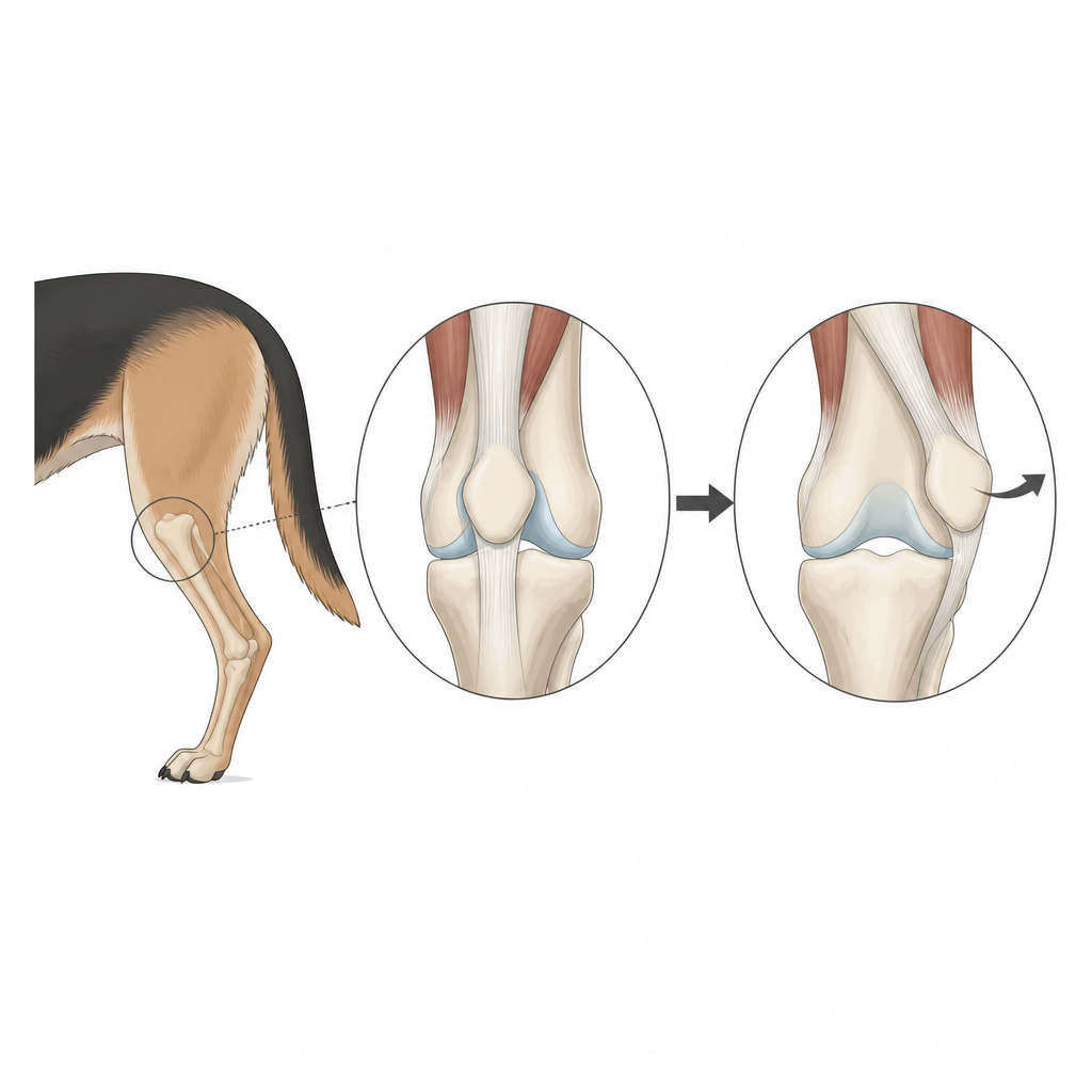
In a healthy knee, the small bone at the front of the joint, the kneecap, runs neatly in a groove on the thigh bone. In dogs with patellar luxation, this kneecap repeatedly jumps out of that groove. The researchers examined 14 affected knees from client owned dogs and compared them with two healthy knees from dogs without joint disease. They focused on two key tissues inside the joint: the shiny, shock absorbing joint surface and the thin capsule that lines and surrounds the joint.
Looking closely at the joint surface
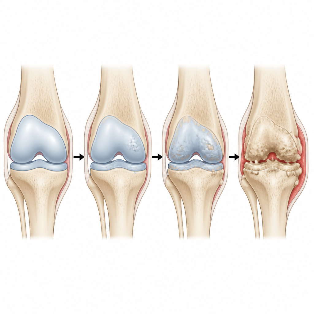
Under the microscope, healthy joint surfaces showed a smooth top layer and well spaced cells that keep cartilage healthy. In dogs with a mildly slipping kneecap, this surface already showed small rough patches and early changes in cell shape. In more severely affected knees, cracks, pits, and areas of clear cell loss appeared, and some cartilage blended abnormally into the underlying bone. The most severe group had many empty spaces where living cells should have been, and the surface was scarred and fibrous, clear signs of advanced wear and tear.
Changes in joint “cushion” chemicals
The team also stained the cartilage to measure proteoglycans, molecules that help cartilage hold water and act as a cushion. Healthy samples took up the stain strongly in all layers, showing rich chemical content. As kneecap slipping became more severe, the stain weakened, especially near the surface, meaning the cartilage was losing some of its cushioning ability. While differences between the mild and moderate groups were not all statistically clear, the most severe group showed noticeably poorer staining than normal joints, pointing to chemical as well as structural damage.
Inflamed joint lining and visible surgical findings
The thin tissue that lines the joint and helps make joint fluid also changed with disease severity. In mildly affected dogs it showed small areas of bleeding and extra blood vessels. In moderate and severe cases, there were many more immune cells, more supporting cells, and in the worst joints even fragments of abnormal bone with signs of bony overgrowth. During surgery, the veterinarians often saw bony spurs and bare patches on the cartilage surface, especially on the lower part of the kneecap and along parts of the groove where it should normally slide.
What this means for dogs and their care
Taken together, the findings show that a slipping kneecap is linked with real, structural harm to the dog’s knee, affecting both the smooth joint surface and the surrounding lining. The worst grades of luxation tended to have the greatest loss of cartilage cells, more disturbed surface structure, and weaker cushioning chemicals, even though the small number of dogs limited how sharply all grades could be separated statistically. For owners and veterinarians, this supports the idea that early diagnosis and well planned surgery, followed by good aftercare, may help limit ongoing joint damage and reduce the risk of later arthritis.
Citation: Sharma, P., Anand, A., Pathak, D. et al. Stifle joint alterations in dogs with patellar luxation. Sci Rep 16, 15810 (2026). https://doi.org/10.1038/s41598-026-44207-y
Keywords: patellar luxation, dog knee, stifle joint, cartilage damage, canine orthopedics