Clear Sky Science · en
Multi-dimensional deep learning–based segmentation and volumetric assessment of sphenoid sinus fluid on postmortem CT in drowning cases
Why this matters beyond the autopsy room
When someone is found dead in water, even experienced forensic doctors can struggle to say with confidence whether the person truly drowned or died from something else beforehand. One subtle clue is fluid that collects in a small air-filled pocket deep behind the nose, called the sphenoid sinus. Measuring that fluid on postmortem CT scans could help, but doing it by hand is slow and depends heavily on who is reading the images. This study explores whether modern computer vision techniques can automatically find and measure that fluid, offering faster and more consistent support for difficult drowning investigations. 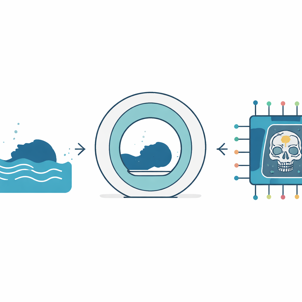
A hidden pocket with important clues
In drowning cases, fluid often seeps into the paranasal sinuses, tiny chambers in the bones of the face. The sphenoid sinus, tucked deep in the skull, is especially interesting because fluid collected there can be tested for microscopic algae and for salt levels that hint at whether the person drowned in fresh or seawater. Earlier work showed that CT scans taken after death can roughly match the fluid volumes later found at autopsy. But to get those numbers, experts have had to outline the fluid region slice by slice on a computer—a tedious task that is hard to standardize and impractical for large numbers of cases.
Teaching computers to spot fluid
The research team turned to a family of image-analysis systems widely used in medical imaging, known as U-Net–style deep learning models. They gathered postmortem CT scans from 165 people whose deaths by drowning had been confirmed at autopsy. An experienced observer manually traced the fluid in the sphenoid sinus to create a reference map for the computers to learn from. The cases were split into training, validation, and testing groups so that the models would be judged on data they had not seen before. The scientists then built three versions of the system: one that looked at individual CT slices (2D), one that considered each slice together with its two neighbors (2.5D), and one that analyzed small three-dimensional blocks of the head in one go (3D).
How well the models performed
All three models were able to mark out sphenoid sinus fluid on the test scans in a way that closely matched the human expert. The 2D and 2.5D approaches were the most reliable at tracing the shape and borders of the fluid, while the 3D version was somewhat less precise and more variable from case to case. When the team converted these outlines into actual fluid volumes, the automatic estimates were within a fraction of a milliliter of the reference values, and the differences were not statistically significant. The predicted and manual volumes moved in lockstep, with very strong correlations across all three models, suggesting that the computer-generated measurements can stand in for labor-intensive hand measurements. 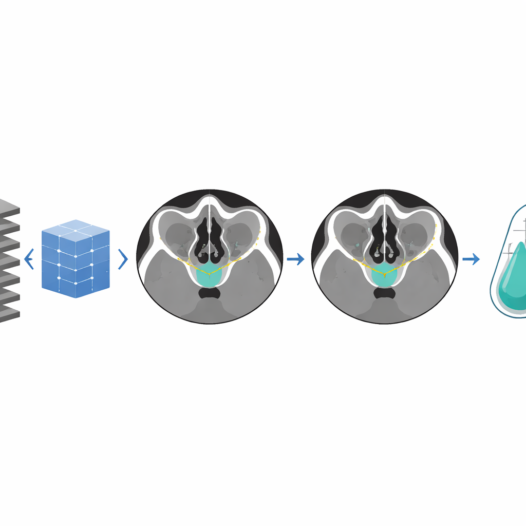
Why a middle-ground approach won
The 2.5D model, which looks at a slice along with its immediate neighbors, offered the best balance between accuracy and practicality. It benefits from some three-dimensional context, helping it distinguish true fluid from nearby bone and soft tissue, yet it avoids the heavy computational demands and instability that can plague full 3D models—especially when the structure of interest is tiny and appears in only a few slices. For busy forensic centers that may need to process many cases with limited computing power, this middle-ground strategy could be the most realistic choice for routine use.
What this means for future death investigations
For non-specialists, the key takeaway is that computers can now reliably find and measure a small pocket of fluid deep inside the head on postmortem CT scans, with performance that closely matches a human expert. The study shows that such automated measurements can be produced quickly and consistently, opening the door to using CT data to support drowning diagnoses even before an autopsy begins. While this single-center work still needs to be tested more widely and extended to other sinuses and to non-drowning deaths, it points toward a future in which advanced image analysis quietly strengthens the evidence behind some of the most challenging questions in forensic medicine.
Citation: Heo, JH., Kim, MJ., Jang, S.J. et al. Multi-dimensional deep learning–based segmentation and volumetric assessment of sphenoid sinus fluid on postmortem CT in drowning cases. Sci Rep 16, 13800 (2026). https://doi.org/10.1038/s41598-026-44094-3
Keywords: drowning diagnosis, postmortem CT, forensic imaging, deep learning, sinus fluid