Clear Sky Science · en
Visual field progression in varying severities of treated patients with primary angle closure glaucoma
Why this eye study matters
Glaucoma is a leading cause of permanent blindness, and one common form, primary angle closure glaucoma, is especially sight threatening in Asia. This study followed hundreds of patients for at least five years to see how quickly their side vision declined and which patients were most at risk. The findings can help doctors tailor care so that people keep useful vision for as long as possible.
Different stages of disease
Not all glaucoma is equally advanced when it is first found. In this work, doctors grouped Chinese patients into three bands based on how much of their visual field was already damaged: mild, moderate, or severe disease. All patients were receiving regular care at a large eye center and had at least five reliable tests of their side vision over time. This allowed the team to track how quickly each person lost function even while under treatment. 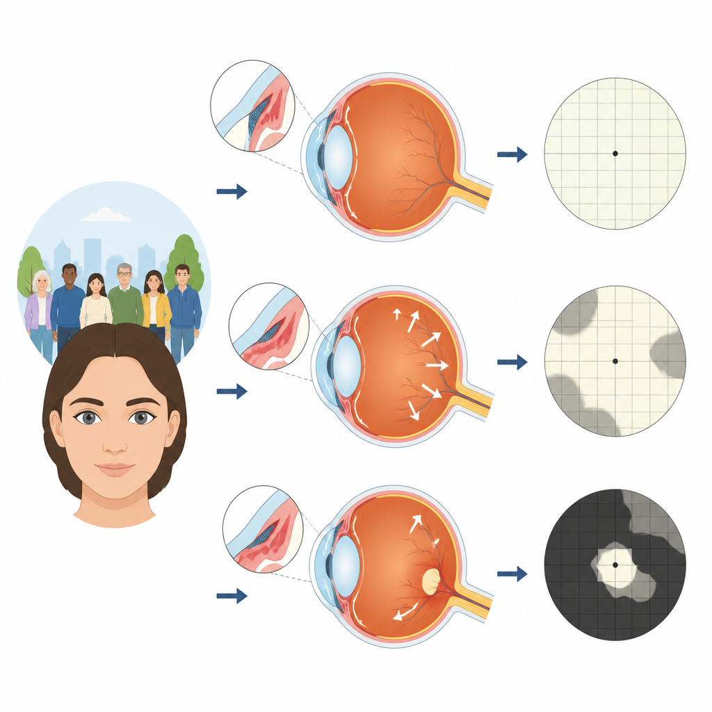
How fast vision was lost
When the researchers compared the three groups, they saw an unexpected pattern. On average, patients with moderate disease lost vision fastest, while those with mild or very severe disease worsened more slowly. This U shaped curve suggests that the middle group sits in a dangerous zone where there is still enough vision to lose quickly, but the damage is already far along. About half of all eyes changed only slowly, a third declined at an intermediate pace, and a smaller group lost vision quickly despite treatment.
What drove change in early and middle stages
The team then examined which clinical features were linked with the speed of change at each stage. In milder disease, eyes with higher pressure inside the eye and more pressure swings from visit to visit tended to lose vision more quickly. Shorter eye length also appeared to offer some protection, hinting at a complex role for eye shape. In moderately affected eyes, greater pressure fluctuation again went hand in hand with faster loss, while features such as poorer starting sharpness of sight were tied to slower change, perhaps because these eyes received more intensive care.
What mattered in advanced disease
In the most advanced group, the picture was different. Eyes that progressed quickly often did not have very high pressures at the start and tended to have wider drainage angles, findings that do not fit the classic story of pressure driven damage alone. The authors suggest that long term injury to the optic nerve and its blood supply may keep harming vision even after pressure is lowered. They also note that standard visual field tests may underestimate further damage once much of the field is already gone. 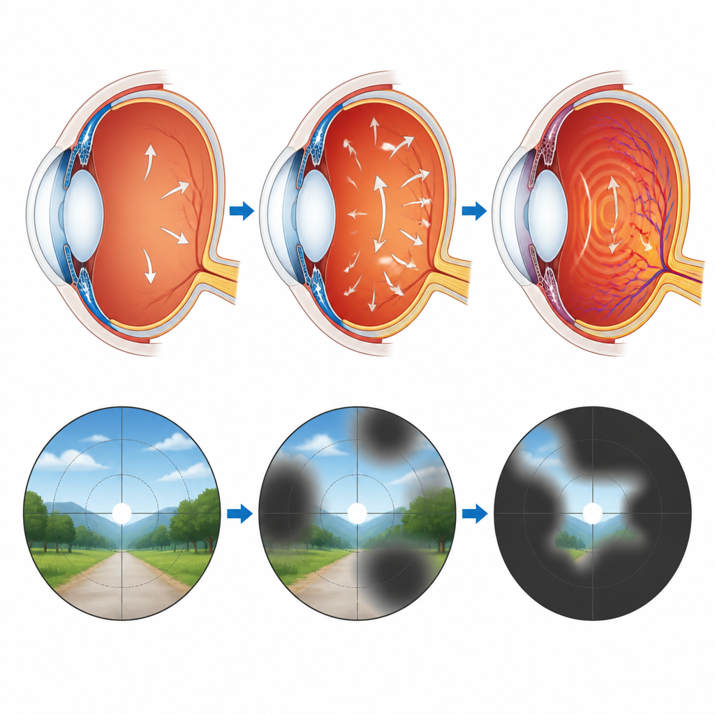
What this means for patients
Overall, the study shows that the risk of vision loss in primary angle closure glaucoma changes with disease stage, and that moderate disease may be the most vulnerable phase. Keeping eye pressure stable, not just low on average, seems especially important early on, while other factors become more important later. The results support close, stage specific monitoring and timely surgery where needed so that people living with this common form of glaucoma can preserve useful sight.
Citation: Tan, S.S., Teng, T.S., Prasad, S. et al. Visual field progression in varying severities of treated patients with primary angle closure glaucoma. Sci Rep 16, 15428 (2026). https://doi.org/10.1038/s41598-026-44049-8
Keywords: glaucoma, primary angle closure, visual field loss, intraocular pressure, eye disease progression