Clear Sky Science · en
Age-dependent alterations of TRPA1 and urocortin 1 signaling in the Edinger–Westphal nucleus in a mouse model of Alzheimer’s disease
Why this brain study matters
Alzheimer’s disease is usually linked to memory loss and brain shrinkage, but less is known about how tiny, deep brain centers shape mood and thinking as the disease unfolds. This study looks at a small cluster of cells in the midbrain of mice that may help connect changes in brain chemistry to problems with stress, emotion, and memory in Alzheimer’s-like disease.
A little-known hub inside the midbrain
Deep in the midbrain lies the Edinger–Westphal nucleus, which actually has two parts. One helps control the eye, but the other, called the centrally projecting Edinger–Westphal nucleus, sends chemical signals widely across the brain. Many of its nerve cells make a stress-related messenger called urocortin 1, which can influence mood, stress adaptation, sleep–wake cycles, pain, eating, and alcohol intake. Earlier work showed that this same cell group is affected in disorders such as Parkinson’s disease and chronic stress, hinting that it might also play a role in the emotional and cognitive problems that accompany Alzheimer’s disease.
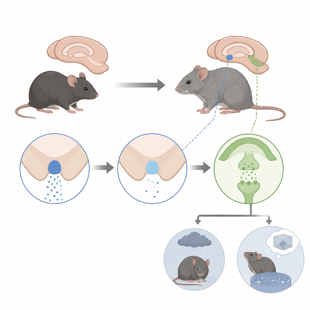
A special sensor and a stress messenger
These midbrain cells also carry a molecular sensor called TRPA1, an ion channel that opens in response to chemical byproducts of inflammation and oxidative stress, both of which are common in aging and Alzheimer’s disease. When TRPA1 opens, calcium flows into the cell and can trigger the release of messengers like urocortin 1. The researchers proposed that, as Alzheimer-like changes accumulate, signals coming from other brain regions such as the hippocampus would alter TRPA1 and urocortin 1 in this nucleus, reshaping stress and mood circuits that connect to areas like the prefrontal cortex, amygdala, hypothalamus, and brainstem.
Following changes across the lifespan of Alzheimer-model mice
To test this idea, the team used male triple transgenic mice that develop key features of Alzheimer’s disease over time and compared them with healthy mice at several ages from young adulthood to old age. In thin brain slices, they counted how many TRPA1 RNA copies each urocortin 1 cell contained and measured how much urocortin 1 peptide had built up inside the cells. In a separate group of animals, they used magnetic resonance spectroscopy to track two chemical markers in the hippocampus, a memory center: N-acetylaspartate, which reflects neuronal integrity, and taurine, a small molecule linked to protection against oxidative stress and to healthy brain metabolism.
What changed in these cells and chemicals
In healthy mice, TRPA1 levels in the midbrain cells started relatively high and then steadily declined with age. In the Alzheimer-model mice, however, TRPA1 expression was already low at young ages and stayed low across life. Urocortin 1 content inside the same cells started out lower in young Alzheimer-model animals than in controls, but then rose with age in both groups and especially in the transgenic mice, before dropping again in the oldest animals. This pattern suggests that urocortin 1 may be produced but not efficiently released, leading to build-up in the cells when TRPA1 signaling is altered. In the hippocampus, taurine relative to creatine fell with age in the Alzheimer-model mice but not in controls, while N-acetylaspartate levels did not show clear age-related or genetic differences, hinting that metabolic stress might precede major neuron loss.
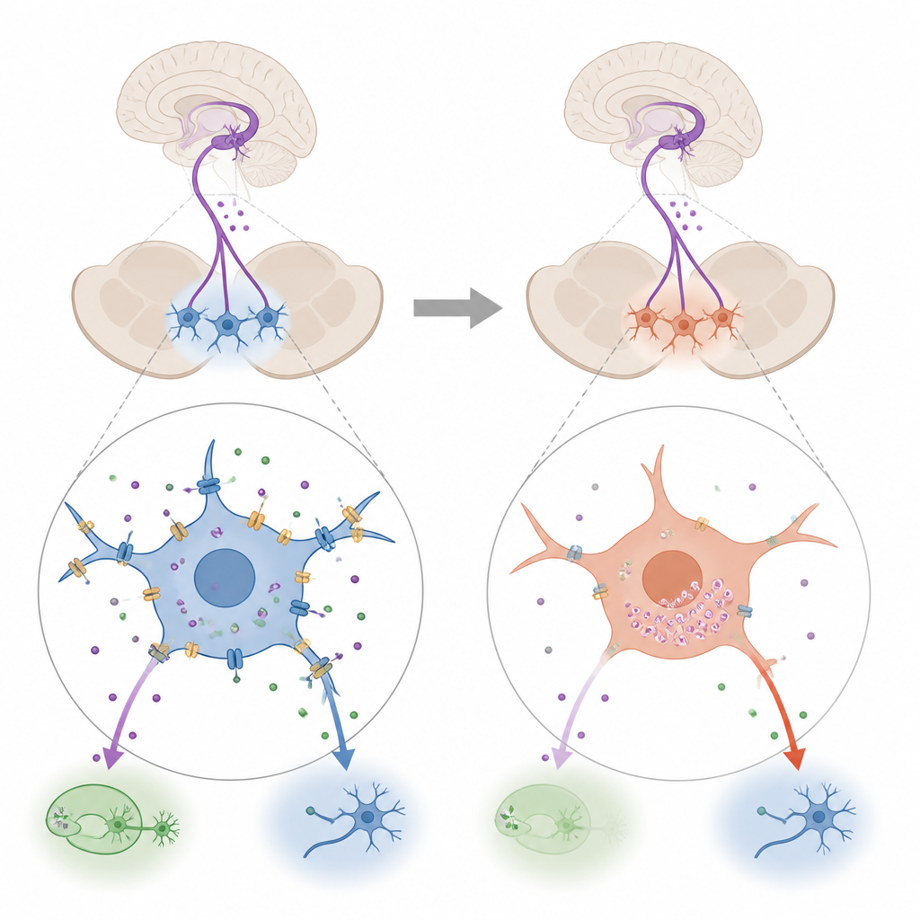
How this may relate to mood and memory
The centrally projecting Edinger–Westphal nucleus sends urocortin 1 signals to brain regions that regulate both mood and memory, including serotonin-producing cells in the dorsal raphe nucleus and circuits involving the amygdala, prefrontal cortex, and hippocampus. The authors suggest that long-lasting inflammatory and oxidative signals in Alzheimer’s disease chronically stimulate TRPA1, which over time may drive the cell to reduce TRPA1 production as a protective response. This, in turn, could disturb urocortin 1 release, upsetting the fine balance of stress and mood regulation and contributing to anxiety, depression, and memory problems seen in aging and Alzheimer’s models.
Take-home message
This work points to a small midbrain hub and its chemical tools, TRPA1 and urocortin 1, as potential links between brain-wide degeneration, stress chemistry, and the emotional and cognitive symptoms of Alzheimer’s disease. Rather than focusing only on memory centers, the study highlights how hidden control nodes and their changing signaling patterns across age might help explain the complex mix of mood and memory changes that people experience as dementia progresses.
Citation: Prókay, A.P., Konkoly, J., Kormos, V. et al. Age-dependent alterations of TRPA1 and urocortin 1 signaling in the Edinger–Westphal nucleus in a mouse model of Alzheimer’s disease. Sci Rep 16, 14829 (2026). https://doi.org/10.1038/s41598-026-44022-5
Keywords: Alzheimer’s disease, Edinger–Westphal nucleus, TRPA1, urocortin 1, hippocampal taurine