Clear Sky Science · en
Region guided mask R-CNN with Haralick ResNet fusion for accurate coronary artery disease detection in computed tomography angiography images
Why clearer heart scans matter
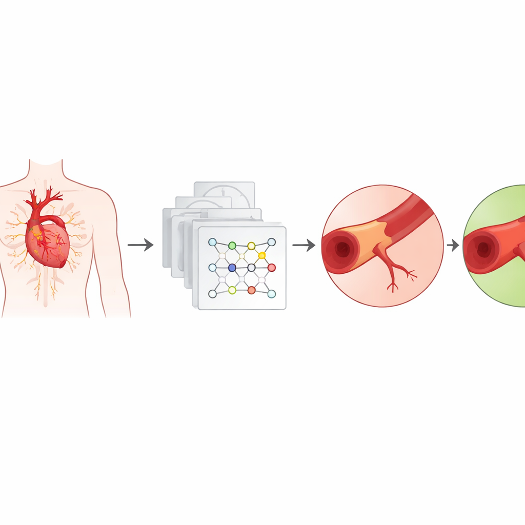
Coronary artery disease is one of the leading causes of death worldwide, and a common way to spot it early is with special CT scans that show the heart’s blood vessels. These 3D images can reveal dangerous narrowings and fatty deposits before they trigger a heart attack. But in real life, the pictures are often fuzzy, crowded with overlapping tissues, and sprinkled with noise. This paper presents a smarter computer system that cleans up those messy images, traces the heart’s vessels more precisely, and flags disease with high reliability, potentially giving doctors a sharper tool for early diagnosis.
The challenge of reading heart pictures
Coronary CT angiography creates detailed maps of the arteries that feed the heart. Yet these images are far from perfect: motion blur from a beating heart, low contrast between vessels and surrounding tissue, and calcium deposits that look similar to nearby structures all make the pictures hard to interpret. Traditional computer methods try to carve out arteries by simple rules, such as picking pixels above a brightness threshold or following edges. These older tools tend to break down when vessels are thin, twisted, or overlapped by other anatomy, which can lead to missed disease or false alarms.
Guided tracing of the heart’s vessels
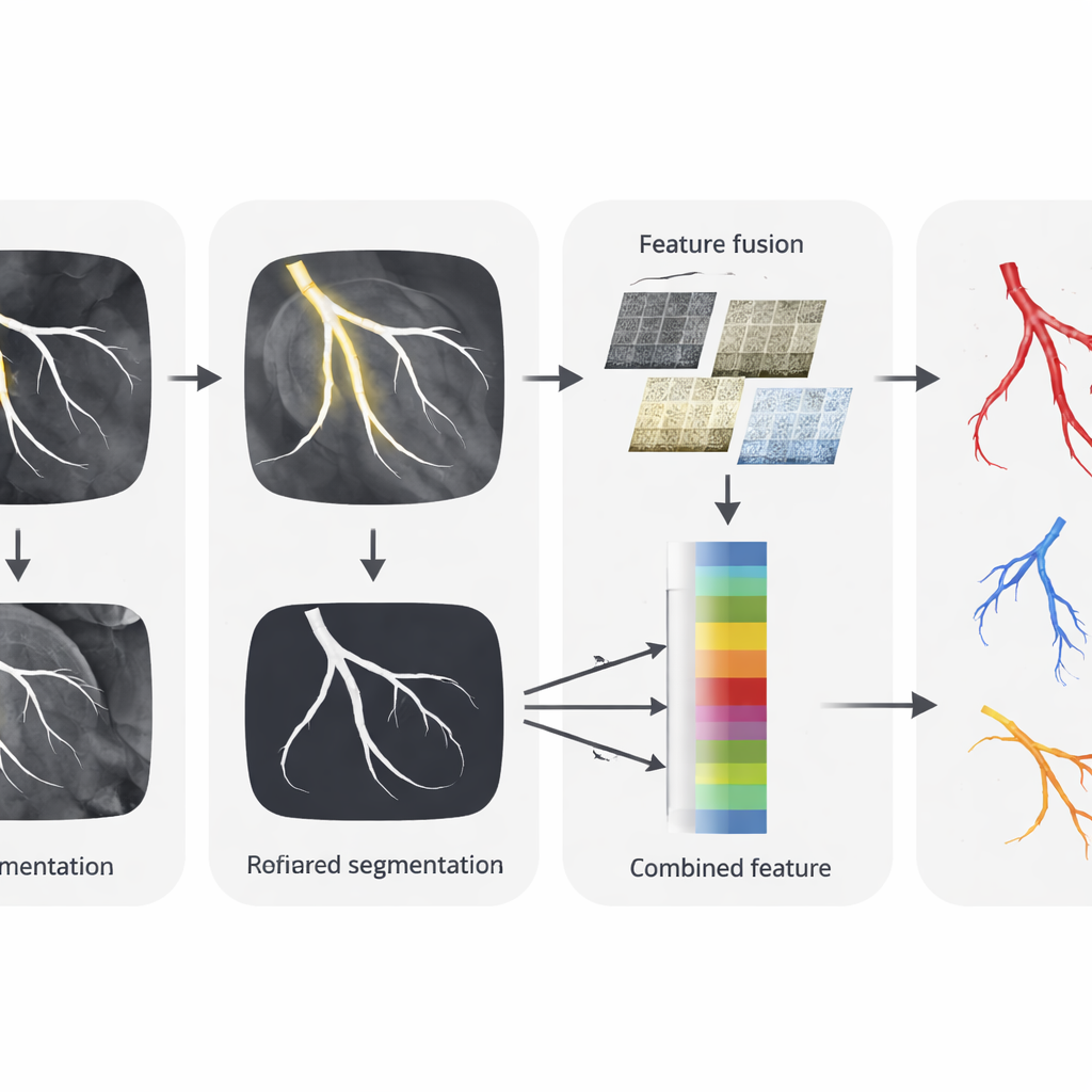
To handle these tricky images, the authors introduce a new way to outline coronary arteries called Region-Guided Mask R-CNN. First, a simple region-growing step marks likely vessel areas, using rough intensity similarities to point out where arteries might be. Then an advanced deep learning model takes over to refine these hints into crisp, object-by-object vessel masks. The system adds a form of visual “attention” that learns to focus on the most relevant regions and ignore clutter, and uses more precise alignment so fine details are not lost. Tested on public heart scan data from 206 patients, this guided tracing achieved higher accuracy and better overlap with expert-drawn vessel outlines than popular segmentation networks such as U-Net and V-Net.
Blending texture and deep learning insight
Spotting disease is not only about drawing vessel borders; it also requires understanding subtle differences inside the artery walls. The researchers build a second component, called Haralick-ResNet Fusion, to capture both fine texture and broader patterns. One part measures classic texture features—how pixel shades repeat, contrast, and blend—while another part, a modern deep network (ResNet), learns complex visual cues directly from the data. These two streams are merged into a single, richer description of each artery region. This fused representation helps the system distinguish normal vessels from those affected by plaque or narrowing, even when visual differences are slight.
From pictures to a clear yes-or-no answer
The final step in the pipeline is a deep convolutional network that takes the fused features and decides whether coronary artery disease is present. This classifier layers many small filters to recognize patterns at different scales, then condenses them into a decision. Using careful data preparation, image normalization, and augmentation, the model reached an overall disease detection accuracy of about 98.3 percent. It also outperformed well-known image classification networks such as VGG-16, ResNet-50, InceptionV3, and EfficientNet-B3 on the same task, while maintaining reasonable computation time and frame rate.
What this means for heart care
Viewed from a layperson’s perspective, this work is about teaching computers to read heart scans more carefully and consistently than current systems. By first tracing the arteries with region-guided attention, then blending classic texture cues with deep learning insight, and finally issuing a confident yes-or-no verdict, the method tackles the main obstacles that have limited automated analysis of coronary CT images. While it still depends on good-quality scans and further clinical validation, the approach suggests that smarter, end-to-end AI tools could soon help radiologists spot risky heart disease earlier and more reliably, potentially guiding treatment before symptoms become life-threatening.
Citation: Revathi, G., Mathew, O.C. Region guided mask R-CNN with Haralick ResNet fusion for accurate coronary artery disease detection in computed tomography angiography images. Sci Rep 16, 12231 (2026). https://doi.org/10.1038/s41598-026-43951-5
Keywords: coronary artery disease, cardiac CT imaging, medical image segmentation, deep learning diagnosis, heart plaque detection