Clear Sky Science · en
Transcranial cisternal port enables repetitive intrathecal delivery in mice
A New Window into the Brain’s Protective Fluid
The brain and spinal cord float in a clear liquid called cerebrospinal fluid, which both cushions them and carries medicines and waste products. Doctors are increasingly trying to treat brain cancers by delivering drugs directly into this fluid, but it has been hard for scientists to study such treatments in mice, whose small size makes repeated access to this space technically difficult. This study introduces a simple, durable “port” in the skull of a mouse that lets researchers safely reach this fluid again and again, opening the door to more realistic testing of future brain therapies.
Why Getting Drugs into the Brain Is So Hard
Many promising drugs for brain diseases never make it to their targets because of natural defenses such as the blood–brain barrier, which tightly controls what can leave the bloodstream. One way around this problem is to inject medicines directly into the fluid-filled space that surrounds the brain and spinal cord. In people, devices like the Ommaya reservoir allow repeated treatments through a small dome under the scalp. In mice, however, most methods allow only single injections or rely on tiny plastic tubes that can clog, shift position, or leak, making experiments slow, imprecise, and difficult to scale up.
Designing a Tiny but Steady Access Port
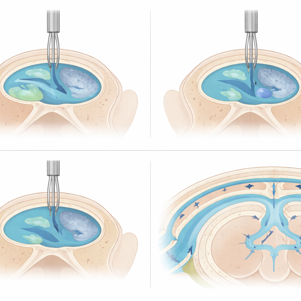
The researchers tackled this challenge by building what they call a Transcranial Cisternal Port, or TCP, sized for the mouse skull. The cisterna magna, a fluid-filled pocket at the back of the brain covered by a thin, see-through membrane, serves as the gateway. Surgeons make a small opening in the skull just above this pocket and create a shallow “seat” in the bone for a short metal tube, or cannula. The cannula is angled so that its tip points directly toward the cisterna magna. Using a microscope, they introduce a very thin wire through the cannula and visually confirm that it appears in the fluid space, then glue the base of the cannula firmly to the skull, close the skin, and cap the tube with a matching plug to keep it open.
How the Port Performs in Practice
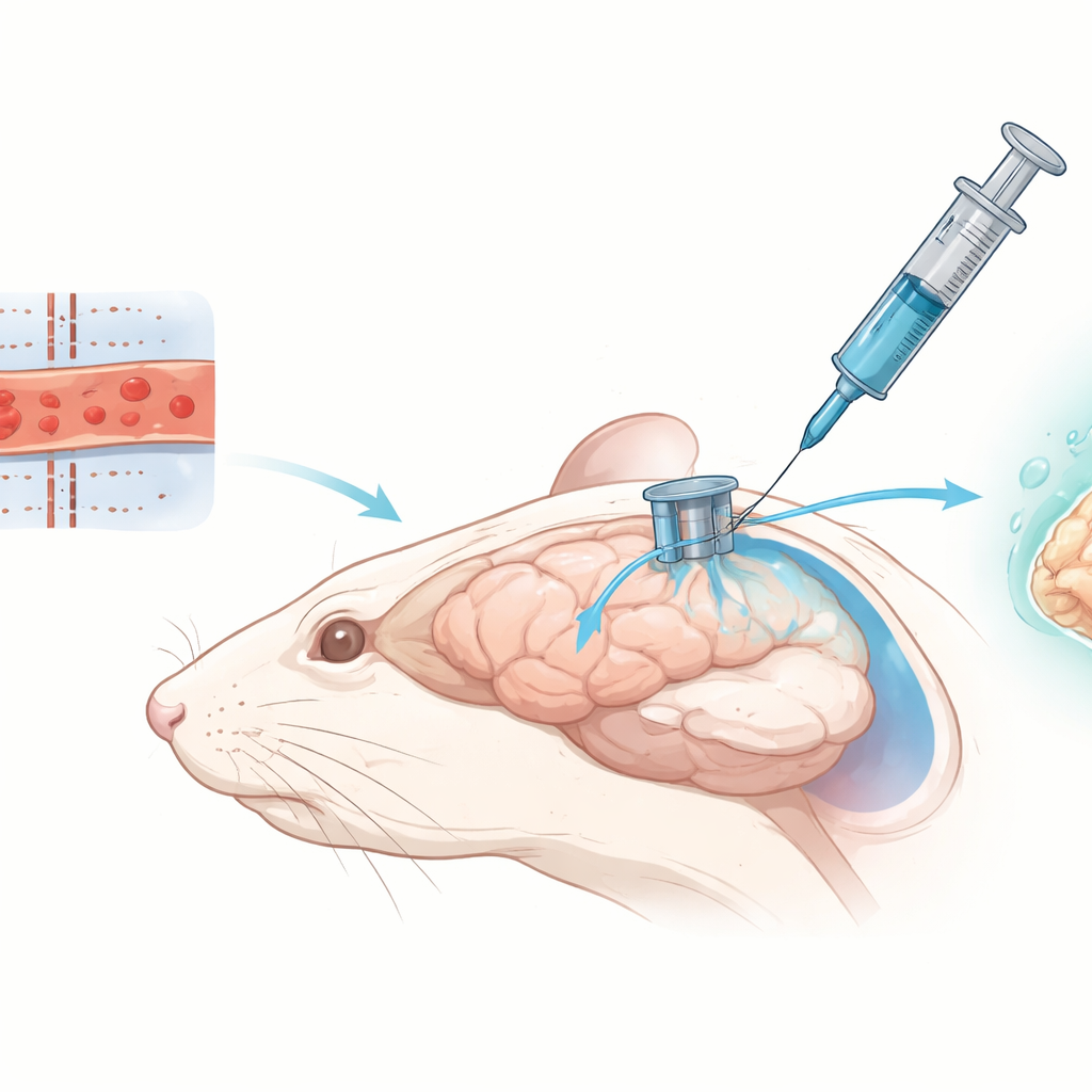
To test whether the port really delivers fluid where intended, the team injected a blue dye through the cannula into the mice. When they examined the brains, they saw dye spreading through the cisterna magna, flowing along fluid channels at the base of the brain, and seeping into the narrow spaces that surround blood vessels within the brain tissue. This pattern matches how cerebrospinal fluid normally circulates, suggesting that medicines delivered through the TCP would likewise reach widespread areas. The team then followed 43 mice for three weeks while using the port repeatedly. All animals recovered normal movement and behavior after surgery, with no signs of leakage, infection, or neurological problems linked to the device itself. After one week, 93 percent of ports remained usable; after two and three weeks, 86 percent were still functioning.
Lessons from Failures and Fine-Tuning
When ports did fail, it was usually for practical, fixable reasons. In four cases, material inside the tube clogged the narrow channel, likely due to sticky components of the injected solutions. In two other mice, a small screw-in plug bonded to the tube when glue used during surgery crept into the threads. Importantly, the rigid metal design allowed surgeons to reopen or replace ports by revisiting the same skull opening, a task that would be much harder with soft plastic tubing buried in muscle. Once the surgical technique was refined, an experienced team could place ports in about ten minutes per mouse, making it feasible to equip dozens of animals for large studies.
What This Means for Future Brain Treatments
To a non-specialist, the key message is that the authors have built a reliable “access hatch” into the mouse’s protective fluid around the brain. This hatch is anchored to bone, can be checked visually during placement, and allows repeated injections over weeks with high success rates and minimal harm to the animal. While clogging remains the main drawback, future versions with slightly wider channels should improve long-term performance. By making it practical to give many rounds of treatment directly into brain fluid, the Transcranial Cisternal Port provides a powerful new tool for testing immunotherapies, cancer drugs, and other advanced treatments in realistic preclinical models—an important step toward safer and more effective therapies for human brain diseases.
Citation: Haupt, B., Turunen, J., Olson, I. et al. Transcranial cisternal port enables repetitive intrathecal delivery in mice. Sci Rep 16, 12905 (2026). https://doi.org/10.1038/s41598-026-43886-x
Keywords: intrathecal drug delivery, cerebrospinal fluid, brain cancer models, mouse neurosurgery, cisterna magna