Clear Sky Science · en
Prognostic impact of renal microcirculatory dysfunction in heart failure assessed by superb microvascular imaging
Why the Heart–Kidney Connection Matters
When the heart begins to fail, it does not struggle alone. The kidneys, which quietly filter our blood around the clock, are deeply affected by changes in blood flow and pressure. This study explores a new ultrasound technique that lets doctors watch tiny blood vessels inside the kidney in real time. By seeing how well these delicate vessels are working, the researchers show that we may be able to predict which patients with heart problems are at higher risk of landing back in the hospital or dying—well before traditional tests raise a red flag. 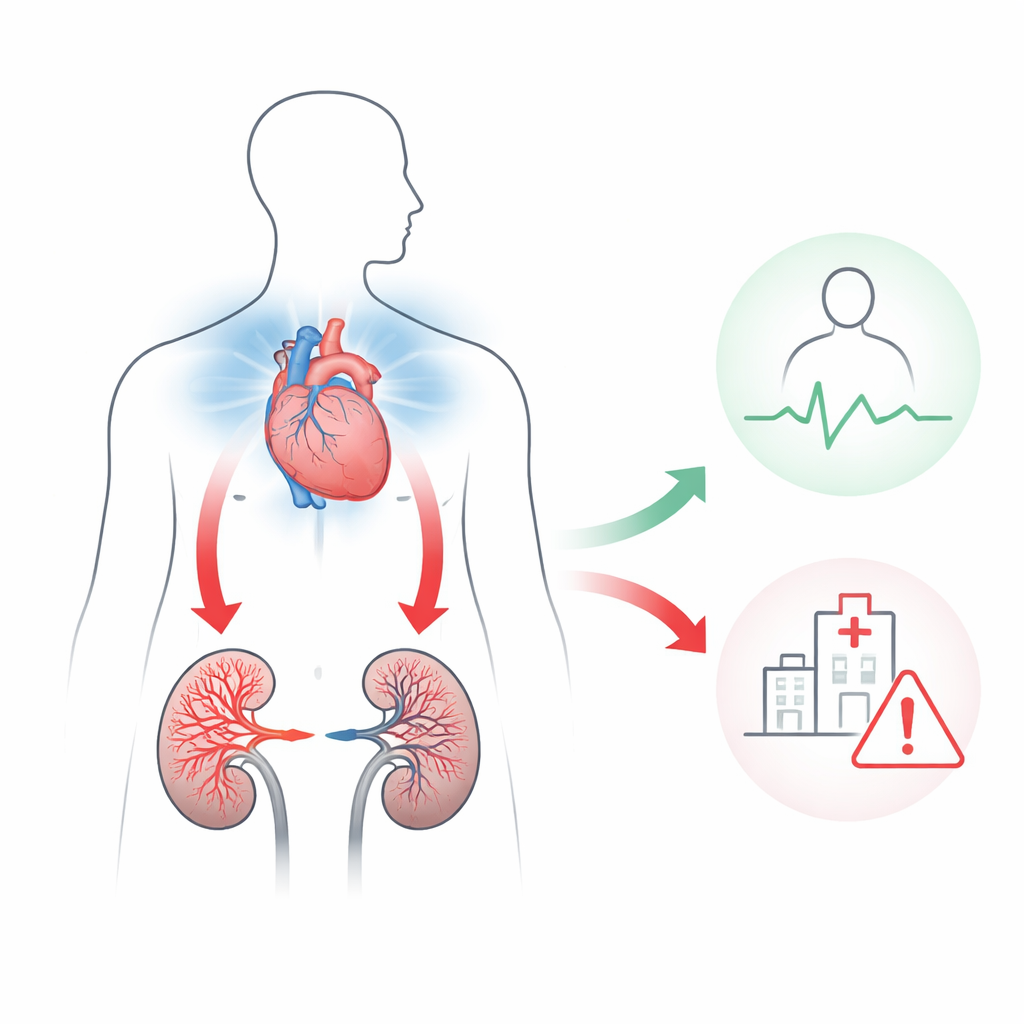
A Closer Look at Tiny Kidney Vessels
The kidneys are packed with a rich network of miniature blood vessels that must stay open and well supplied for the organs to do their job. In heart failure, the heart’s reduced pumping power and rising pressures in the veins can squeeze these vessels, slowing or interrupting flow. The team used a specialized form of ultrasound, called superb microvascular imaging, that is sensitive enough to detect slow, fine blood movements without contrast dyes. From these images, they calculated a “vascular index,” which reflects how much of the kidney area is filled with blood flow at any given moment.
Measuring Blood Flow Over Each Heartbeat
Instead of taking just a single snapshot, the researchers tracked how the vascular index rose and fell during each heartbeat. They defined a maximum value, a minimum value, and how much the signal swung between the two over one cardiac cycle. This swing, called the intrarenal perfusion index, hints at how steady or choppy the blood supply is: more fluctuation suggests that kidney vessels are being stressed by changing pressures. In earlier work, the same group had linked these image-based measures to pressures inside the heart’s right side, but it was not yet clear whether they could forecast future health problems.
Following Patients Over Time
The study reviewed 78 people who underwent kidney scanning with this technique, most of whom already had chronic heart failure. Over an average of about a year and a half, 13 patients died or were unexpectedly hospitalized for worsening heart failure. Those who suffered these serious outcomes tended to have lower maximum and minimum vascular index values—meaning their kidneys showed less overall tiny-vessel blood flow—and greater swings over each heartbeat. Statistical models confirmed that these three image measures were strongly linked with risk, even after accounting for age, blood tests of kidney function, estimated pressure in the central veins, and other ultrasound markers traditionally used in heart care.
Seeing More Than Standard Tests Can Show
Doctors usually judge fluid balance and kidney health in heart failure using blood work, overall blood pressure, and rough estimates of pressure in large veins. Yet these broad measures can be misleading. A person can have “normal” central venous pressure while the small vessels inside the kidneys are still congested or starved of blood. In this study, many patients had seemingly low central pressure but already showed disturbed flow patterns in the kidney veins. The new imaging measures, which visualize actual microcirculation, outperformed standard markers such as creatinine in predicting bad outcomes. This suggests that watching the fine details of kidney blood flow may give a more honest picture of how the heart and kidneys are coping together. 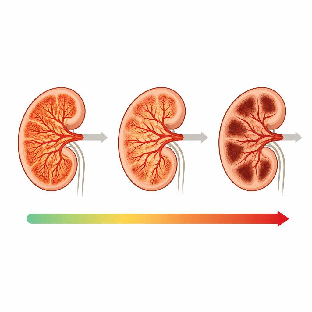
What This Could Mean for Patients
The authors conclude that superb microvascular imaging of the kidney offers a promising, noninvasive way to gauge risk in people with heart failure. By revealing how much blood truly reaches the kidney’s smallest vessels—and how stable that flow is—these scans may help clinicians recognize dangerous congestion or poor perfusion earlier than today’s methods allow. If confirmed in larger, forward-looking studies, this approach could guide more personalized treatment, helping doctors fine-tune fluid removal and medications to protect both the heart and kidneys, and ultimately reduce repeat hospital stays and deaths.
Citation: Kayama, K., Kikuchi, S., Sugimoto, T. et al. Prognostic impact of renal microcirculatory dysfunction in heart failure assessed by superb microvascular imaging. Sci Rep 16, 14055 (2026). https://doi.org/10.1038/s41598-026-43872-3
Keywords: heart failure, kidney blood flow, ultrasound imaging, cardiorenal syndrome, risk prediction