Clear Sky Science · en
Molecular and spatial heterogeneity of macrophage like vascular smooth muscle cells in abdominal aortic aneurysms associated with intraluminal thrombus
Why this matters for artery health
Abdominal aortic aneurysms are silent bulges in the body’s main artery that can burst without warning, often with fatal results. Doctors have long noticed that many of these weak spots are lined with a jelly-like blood clot, called intraluminal thrombus, but have struggled to tell whether this clot is helpful or harmful. This study peels back the layers of the aneurysm wall to show how that clot reshapes nearby cells and stokes inflammation, offering clues to why some aneurysms suddenly fail — and how we might one day stop them.
Hidden changes in the artery wall
The aorta’s middle layer is normally built from smooth muscle cells that behave like steady “contractile” workers, keeping the vessel strong and able to adjust its diameter. In aneurysms, many of these cells abandon their quiet, structural role and adopt new identities. Using advanced staining techniques on human tissue, the researchers found a striking increase in cells that carry both smooth muscle and immune-cell markers in aneurysms that contain a wall-adherent clot. These hybrid cells, called macrophage-like vascular smooth muscle cells, were far more common when thrombus was present, hinting that the cloted environment pushes cells toward a more inflammatory, potentially destructive state. 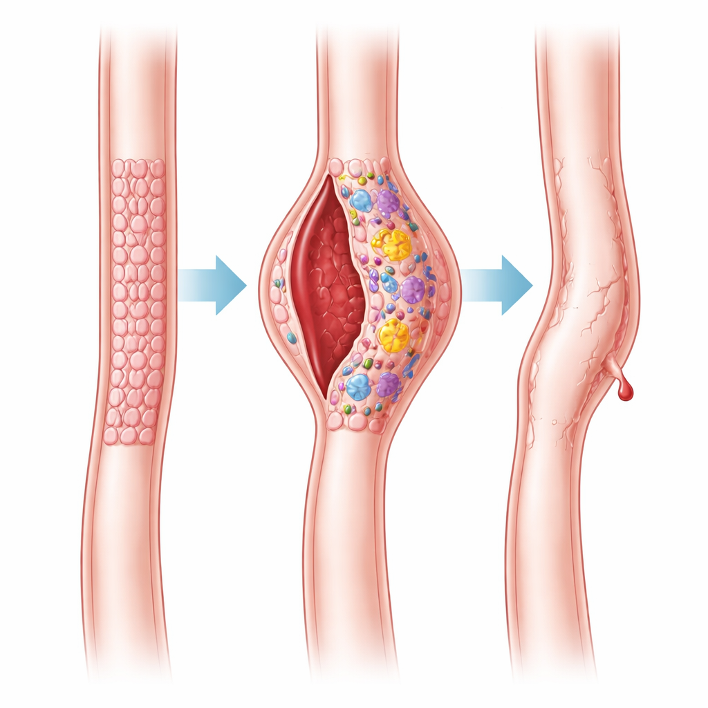
Mapping the molecular neighborhood
To understand what these hybrid cells are doing, the team turned to digital spatial profiling, a technology that reads out thousands of genes from precisely chosen microscopic regions while preserving their location in the tissue. Comparing macrophage-like cells from aneurysms with and without thrombus, they uncovered hundreds of genes that were switched on more strongly when clot was present. Many of these genes are tied to inflammation, tissue breakdown, and stress responses, including well-known inflammatory messengers such as interleukin-6 (IL-6) and interleukin-1β (IL-1β). Pathway analysis showed that signaling routes linked to cell survival, scarring, and immune activation — including NF-κB, JAK/STAT, and PI3K/Akt — were all more active in clot-associated regions.
Immune cells crowd the danger zone
Gene signatures can also reveal what kinds of immune cells are likely lurking nearby. Using a computational tool that decomposes gene patterns into estimated cell types, the researchers found that areas rich in macrophage-like smooth muscle cells also contained higher predicted levels of neutrophils, certain macrophages, and naïve B cells when thrombus was present. The expression of IL-6 and IL-1β strongly tracked with neutrophil-related signals and markers of other activated immune cells, suggesting a feedback loop: clot attracts and activates immune cells; those cells and their products, in turn, encourage smooth muscle cells to behave more like inflammatory scavengers, amplifying local damage.
Neutrophil webs push cells over the edge
To move beyond correlations in human tissue, the team recreated part of this environment in the lab. They isolated neutrophils from healthy donors and triggered them to release fragile, net-like structures known as neutrophil extracellular traps. When smooth muscle cells were exposed to these webs, they lost markers of their contractile identity, gained genes associated with a more synthetic, migratory, and immune-like state, and began secreting inflammatory proteins including IL-1β, IL-6, and IL-18. The cells also became more mobile and showed activation of the NF-κB pathway — a central switch in inflammation. Blocking key components of this pathway or a related sensor, NLRP3, blunted these changes, implying that neutrophil traps drive smooth muscle reprogramming through specific molecular circuits. 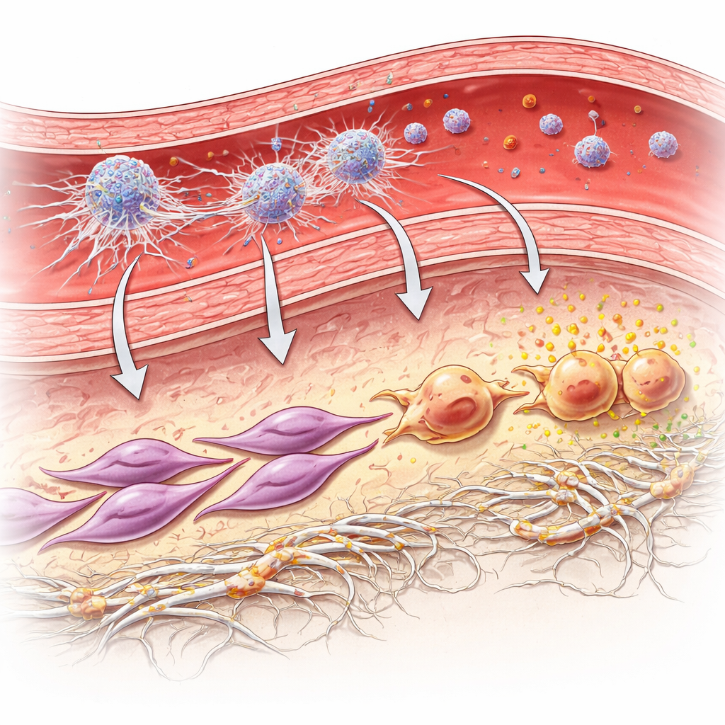
What this means for patients
Taken together, the findings support a picture in which the mural clot in an abdominal aortic aneurysm is not just a passive bystander. Instead, it forms a niche rich in activated neutrophils and their extracellular traps, which nudge structural smooth muscle cells toward a macrophage-like, inflammatory role. These transformed cells then help sustain a cycle of inflammation and tissue breakdown that may weaken the artery wall and promote growth or rupture of the aneurysm. While more work in larger groups of patients and animal models is needed, the study points to new possibilities: therapies that limit neutrophil traps, dampen key signaling pathways like NF-κB or IL-6/IL-1β, or specifically target macrophage-like smooth muscle cells could one day help stabilize aneurysms and reduce the risk of catastrophic rupture.
Citation: Ma, X., Liang, B., Lu, Q. et al. Molecular and spatial heterogeneity of macrophage like vascular smooth muscle cells in abdominal aortic aneurysms associated with intraluminal thrombus. Sci Rep 16, 13654 (2026). https://doi.org/10.1038/s41598-026-43807-y
Keywords: abdominal aortic aneurysm, intraluminal thrombus, vascular smooth muscle cells, neutrophil extracellular traps, vascular inflammation