Clear Sky Science · en
A Machine learning pipeline to investigate tissue ingrowth in cerebral aneurysms using preclinical animal models
Why tiny bulges in brain arteries matter
Cerebral aneurysms are small, balloon-like bulges in brain arteries that can burst without warning, causing stroke or death. One common, less invasive treatment is to pack the bulge with soft metal coils so blood clots and new tissue slowly seals the weak spot from the inside. But doctors and researchers still struggle to measure how well that healing tissue actually fills the aneurysm. The paper behind this summary uses artificial intelligence (AI) to turn high‑magnification microscope images into precise, repeatable measurements of that hidden healing process, with the long‑term goal of helping tailor safer, more durable treatments for patients.
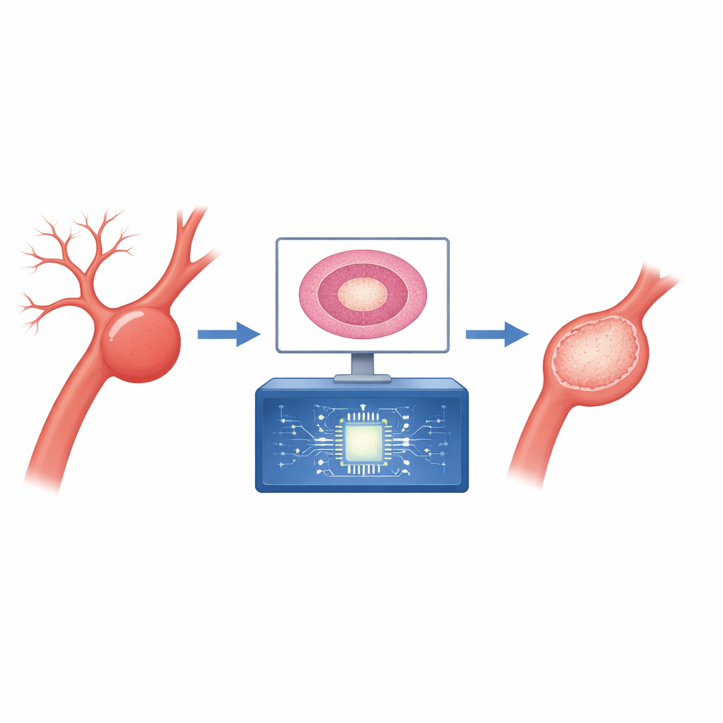
From delicate brain vessels to lab models
In the clinic, aneurysms are treated either by open surgery with a metal clip placed across the neck of the bulge or by endovascular coiling, where tiny coils are threaded up through blood vessels and deployed inside the aneurysm. Coiling is less traumatic and can act like scaffolding for new tissue to grow, but up to a quarter of coiled aneurysms eventually recur and need another procedure. Animal models offer a way to study why some aneurysms heal better than others. In this study, researchers used mice in which carotid artery aneurysms were created and then treated with platinum coils, some coated with molecules designed to encourage tissue repair. Thin, stained slices of the aneurysm region were imaged under the microscope, capturing rich detail about blood clots, connective tissue, and remodeling inside the sac.
Turning microscope pictures into usable numbers
Traditionally, experts inspect these tissue slides by eye and estimate how much new tissue has filled the aneurysm cavity. That approach is slow, subjective, and hard to standardize across labs. The authors designed a machine learning pipeline to automate two key tasks: outlining the aneurysm sac and identifying the areas where new tissue has grown in. They used a modern image‑analysis network called Unet++, originally developed for medical image segmentation, and adapted it to work on high‑resolution histology images. Before training, images were resized, normalized, and augmented with rotations, flips, and scale changes to help the model handle natural variation. Instead of spitting out a simple black‑and‑white mask, the network first produced smooth grayscale “heatmaps” of how likely each pixel belonged to the sac, which were then sharpened using established thresholding methods from image processing.
How well the AI sees what experts see
Once the aneurysm borders were defined, the system measured what fraction of that area was occupied by ingrown tissue. Across 64 images evaluated with ten‑fold cross‑validation, the AI’s sac outlines and ingrowth regions overlapped expert‑drawn regions extremely well, with overlap scores in the mid‑90 percent range and near‑perfect performance on standard classification curves. When the researchers compared the AI’s tissue‑ingrowth measurements to human “ground truth” values, the points lined up closely along the ideal line, showing that the automated method closely tracked expert judgment. To test whether the AI was at least as consistent as trained human helpers, four blinded raters and the model all scored a separate set of images. Agreement with the expert neurosurgeon was highest for the AI, suggesting that the algorithm provides expert‑level consistency, free of fatigue or personal bias.
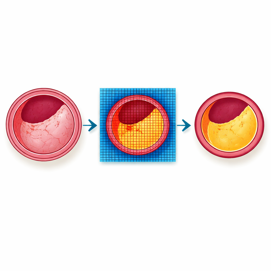
A tool built for non‑experts to use
Recognizing that sophisticated code alone will not transform practice, the team wrapped their pipeline inside a cloud‑based graphical interface that runs in a web browser. Users can upload new aneurysm slides, run the pretrained model, and immediately see colored overlays of the sac and ingrown tissue on top of the original image. The interface also reports simple geometric measurements such as total sac area and ingrowth percentage and allows users to refine boundaries if needed. Because the platform is built on an open‑source digital slide system, other researchers can plug the tool into their own workflows, fostering standardized, shareable measures of aneurysm healing in preclinical studies.
What this means for future aneurysm care
For a layperson, the core message is that this research teaches computers to “measure the scar” inside a treated brain aneurysm with the same reliability as an expert specialist. By replacing rough visual estimates with objective numbers, scientists can more accurately compare coils, drug coatings, and other pro‑healing strategies in animal models before they reach patients. Although the current work is limited to mouse arteries and a single type of staining, it offers a blueprint for how AI can turn complex microscope images into standardized scores of biological stability. In the long run, such tools could help design treatments that make aneurysm repair more complete and durable, lowering the chance that a dangerous bulge in a brain artery ever returns.
Citation: Afsari, F., Ansari, I., Martinez, M.E. et al. A Machine learning pipeline to investigate tissue ingrowth in cerebral aneurysms using preclinical animal models. Sci Rep 16, 13352 (2026). https://doi.org/10.1038/s41598-026-43798-w
Keywords: cerebral aneurysm, endovascular coiling, tissue ingrowth, histology AI, image segmentation