Clear Sky Science · en
Identification of biomarkers associated with endoplasmic reticulum stress-related cell death in osteoporosis based on bulk and single-cell transcriptomic analyses and experimental validation
Why Weak Bones Start Inside Stressed Cells
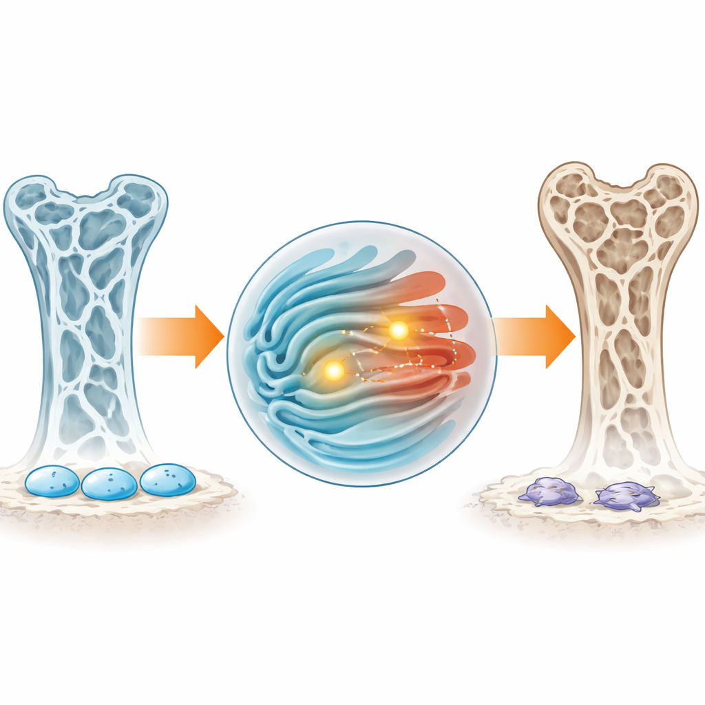
Osteoporosis is best known for thinning bones and unexpected fractures, especially in older adults. But this study looks beneath the X‑ray, into the microscopic machinery inside bone‑related cells. The authors show how a kind of cellular "stress" response and cell death are tied to osteoporosis, and they pinpoint two molecular signals that could help doctors detect risk earlier and guide future treatments.
Looking for Clues in Blood and Bone
Instead of starting from bone scans alone, the researchers examined gene activity patterns in blood cells that can develop into bone‑resorbing cells, and in bone marrow cells taken directly from a patient with osteoporosis. By comparing people with low and high bone density, they narrowed down hundreds of altered genes to those linked both to a key cellular stress system, located in a structure called the endoplasmic reticulum, and to various forms of programmed cell death. They used several machine‑learning methods to sift these candidates, searching for signals that most reliably distinguished osteoporosis from healthy bone status.
Two Molecular Warning Lights
From this extensive screening, two genes stood out: CAMKK2 and DAPK3. Both were consistently less active in people with osteoporosis across multiple datasets, and this pattern was confirmed in blood samples using laboratory tests that directly measure gene activity. When the researchers built a simple prediction tool that combines both markers, it estimated osteoporosis risk with good accuracy in two independent groups of patients. In plain terms, these two molecular warning lights, when dimmed, appear to mark a body that is drifting toward weaker bones.
Stress Signals, Immune Cells, and Possible Medicines
The study also explored what these markers might be doing. When the team looked at broader networks of genes that rise and fall together, CAMKK2 and DAPK3 were linked to pathways that govern how cells respond to inflammatory messengers, how blood vessels form, and how nerve‑like connections adapt over time. They also noticed that people with osteoporosis had fewer of certain immune cells, including activated dendritic cells, and that both markers were modestly tied to the presence of these cells. Computer modeling suggested that existing drugs, such as vitamin D–like compounds that enhance calcium handling, and a hormone‑related medicine once tested for bone loss, might latch onto the proteins made by these genes in a stable way, hinting at new ways to fine‑tune treatment—though this remains speculative.
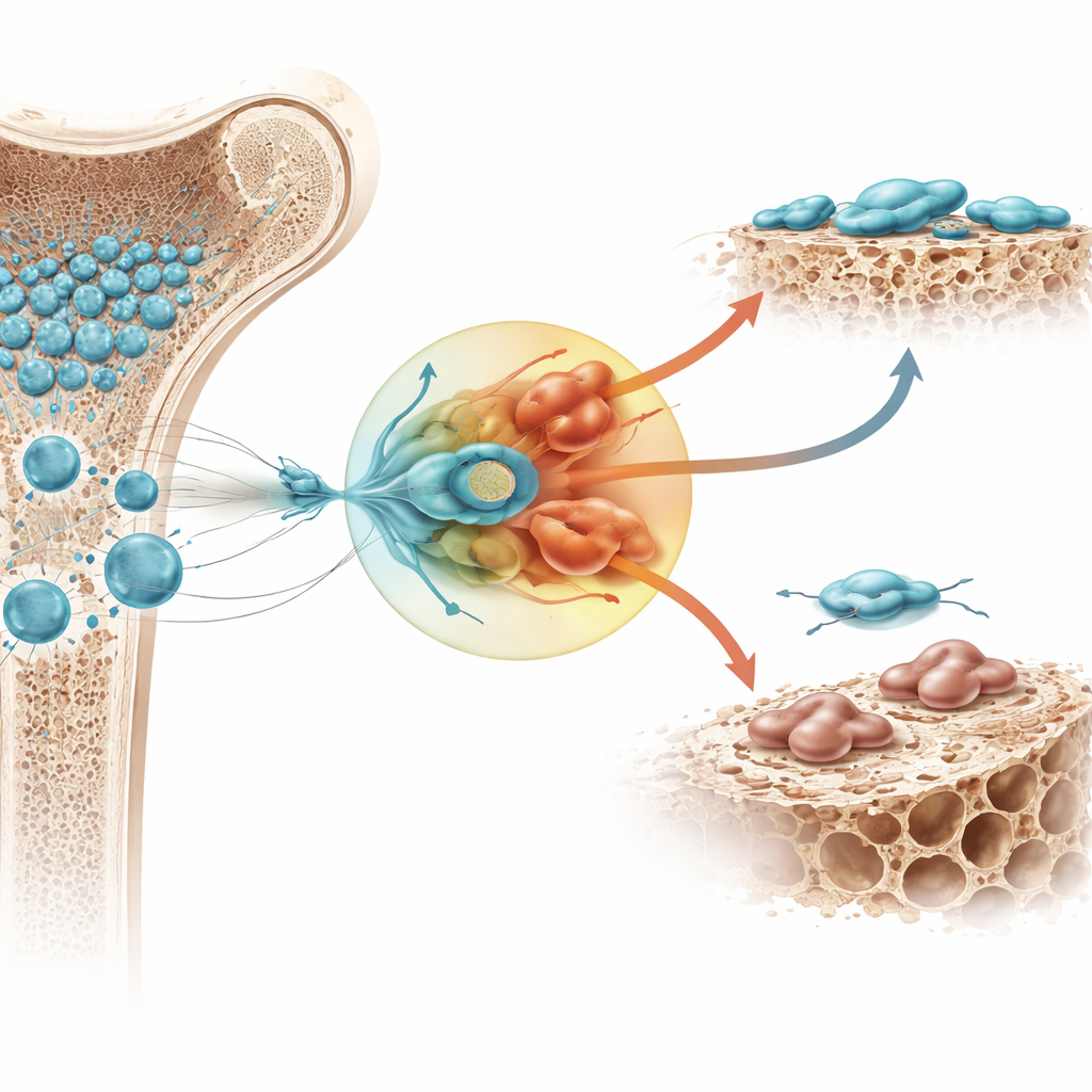
The Role of Stem Cells Inside Bone
To see how all of this plays out at the single‑cell level, the researchers turned to high‑resolution analyses of bone marrow. There, they focused on bone marrow–derived mesenchymal stem cells, a versatile group that can mature into bone‑building cells. These stem cells showed relatively high levels of the two markers and sat at the center of a dense web of communication with nearby immune cells. When the scientists reconstructed the step‑by‑step path as these stem cells matured, they saw that CAMKK2 and DAPK3 activity rose and fell at specific stages, tracking with genes involved in bone formation, cellular stress responses, and controlled cell death. Other pathways that help repair DNA damage or keep cellular ions balanced appeared disturbed in these stem cells, potentially locking them into a stressed state that favors cell loss over healthy bone building.
What This Means for People with Osteoporosis
Taken together, this work suggests that osteoporosis is not just a matter of bone wearing away, but of bone‑forming stem cells trapped in a harmful stress loop that pushes them toward dysfunction and death. CAMKK2 and DAPK3 emerge as key dials in this loop: when they are turned down, the internal stress of the cell and its conversations with immune neighbors may shift in ways that ultimately thin the skeleton. While more studies, larger patient groups, and direct tests in animals are needed, these findings point toward new blood‑based markers for earlier diagnosis and new strategies aimed at calming cellular stress in bone marrow stem cells to preserve bone strength.
Citation: Xia, Y., Peng, Z., Zhao, L. et al. Identification of biomarkers associated with endoplasmic reticulum stress-related cell death in osteoporosis based on bulk and single-cell transcriptomic analyses and experimental validation. Sci Rep 16, 10631 (2026). https://doi.org/10.1038/s41598-026-43744-w
Keywords: osteoporosis, bone marrow stem cells, cell stress, biomarkers, single-cell analysis