Clear Sky Science · en
Nature versus nurture: genetic background and media composition shape endothelial cell transcriptomes in vitro
Why the Same Lab Test Can Give Different Answers
When scientists test how blood vessel cells respond to new drugs or to inflammation, they often use the same kind of cells and similar equipment—yet their results can disagree sharply. This study asks a deceptively simple question with big consequences for medicine: when experiments on human blood vessel cells disagree, is it mostly because people are genetically different, or because the cells are being fed different “soups” of nutrients and growth factors in the lab?
Blood Vessel Cells as a Window into Health
The cells lining our blood vessels, called endothelial cells, help control blood pressure, allow nutrients to pass into tissues, and guide immune cells to sites of injury or infection. Because they touch everything that travels in the bloodstream, they are a prime target for many drugs and a key player in diseases such as atherosclerosis and diabetes. Researchers often turn to human umbilical vein endothelial cells, taken from leftover umbilical cords after birth, as a convenient stand-in for the body’s vessel lining. These cells are widely used because they are relatively easy to obtain and are less shaped by a person’s lifetime of diet, pollution, and illness.
Two Suspects: Genes and the Growth Medium
Despite their popularity, experiments with these cells frequently produce conflicting results—even when teams seem to be studying the same question. The authors focused on two major suspects. One is the natural genetic variation between babies from whom the umbilical cords are collected. The other is the liquid growth medium that bathes the cells in the dish, supplying them with nutrients, blood proteins, and powerful growth signals. Different labs use different recipes, ranging from very rich mixtures that push cells to keep dividing, to leaner blends that keep them in a calmer, more resting state.
A Systematic Look at the Cells’ Inner Workings
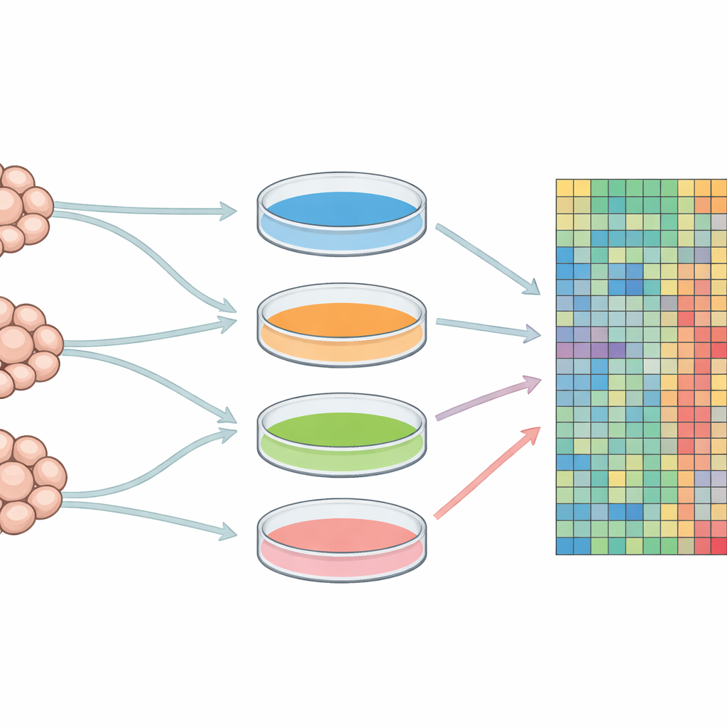
To tease apart these effects, the team grew endothelial cells from three different newborns in four distinct culture media that differed in serum level and added growth factors. They then measured the activity of nearly 14,000 genes in each condition using a microarray, a technology that reads out which genes are turned up or down across the entire genome. By applying several statistical tools, including clustering, correlation analysis, and principal component analysis, they asked which factor—donor identity or medium recipe—did a better job of explaining the patterns in gene activity across all samples.
The Growth Medium Takes the Lead
Across multiple layers of analysis, the message was consistent: the type of growth medium dominated the cells’ genetic behavior. When the researchers grouped samples based on overall gene activity, cells tended to cluster with others grown in the same medium, regardless of which baby they came from. Out of more than two thousand genes that changed strongly in at least one comparison, roughly twice as many were altered by switching media as by switching donors. A commonly used, growth factor–rich medium stood out as especially different from the others, while two media with more modest supplements produced fairly similar gene patterns. Although donor genetics still mattered—hundreds of genes did vary between individuals—the culture conditions had the larger overall impact.
What Changes Mean for Cell Behavior
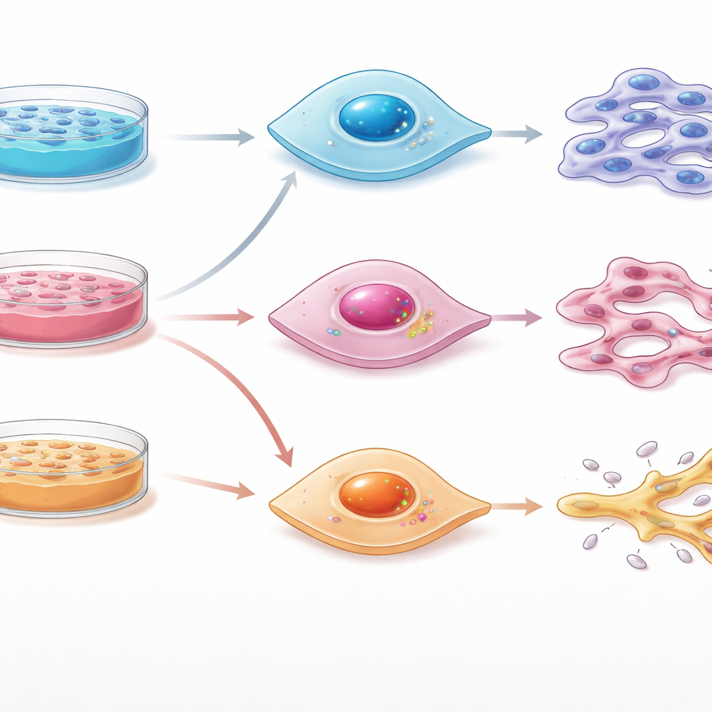
The team then asked what these gene shifts might mean for how the cells actually behave. They grouped genes into everyday functions of vessel-lining cells, such as cell division, formation of new vessel branches, stickiness to circulating cells, leakiness of the vessel wall, and participation in blood clotting and inflammation. Here again, the medium largely set the tone. Richer media strongly boosted gene programs linked to cell division, matching lab experience that they are best for expanding cell numbers. Leaner media favored a calmer, more stable state suited to tests of barrier function. Certain media also sharpened gene patterns related to inflammation, adhesion, and clotting, suggesting that simply changing the recipe in the dish can make cells look more or less “diseased” on the molecular level.
Why This Matters for Reliable Science
For a lay reader, the key takeaway is that what cells are fed in the lab can shape their inner workings even more than who they came from. This means that two studies using “the same” endothelial cells may in fact be probing quite different cellular states if their culture media differ. The authors argue that, alongside using cells from multiple donors, scientists must treat the choice and reporting of growth medium as a central design decision, not a background detail. Doing so could reduce some of the puzzling contradictions in vascular biology and drug testing—and bring us closer to lab results that more reliably predict what will happen inside the human body.
Citation: Demeter, F., Debreczeni, M.L., Németh, Z. et al. Nature versus nurture: genetic background and media composition shape endothelial cell transcriptomes in vitro. Sci Rep 16, 13621 (2026). https://doi.org/10.1038/s41598-026-43732-0
Keywords: endothelial cells, cell culture media, gene expression, experimental reproducibility, HUVEC