Clear Sky Science · en
Menstrual blood-derived stem cell exosomes improve ovarian function in chemotherapy-Induced POF rats via apoptosis regulation
Why this research matters
Many young women who undergo chemotherapy for cancer later discover that their ovaries have been badly damaged, leaving them with irregular periods, early menopause, and difficulty having children. Today’s treatments can ease symptoms but rarely bring the ovaries back to full health. This study in rats explores a hopeful new, cell-free approach: tiny biological packages, called exosomes, collected from stem cells found in menstrual blood, used to help injured ovaries recover and possibly preserve fertility.
A problem beyond surviving cancer
Premature ovarian failure, now often called premature ovarian insufficiency, occurs when the ovaries stop working properly before age 40. It affects hormones, bone health, and emotional well-being, and it can end a woman’s chance of having children with her own eggs. Chemotherapy, while lifesaving, is a major cause because it harms the delicate cells and structures inside the ovary. Standard hormone therapy can replace missing hormones but does not rebuild the ovary itself. Donor eggs are one solution for pregnancy, but they are scarce and do not restore a woman’s own reproductive function, motivating the search for true regenerative treatments.
Turning menstrual blood into a healing resource
Mesenchymal stem cells are a versatile type of cell being tested for many diseases, including ovarian damage. However, transplanting whole stem cells raises safety and practical concerns, such as immune reactions and the risk of clots. Much of their benefit actually comes from exosomes—nano-sized bubbles they release, carrying proteins and genetic signals that can shift how nearby cells behave. Menstrual blood is an especially attractive source for these stem cells: it is easy to collect regularly, does not require surgery, and can provide large, consistent batches of exosomes. In this study, researchers purified exosomes from menstrual blood–derived stem cells (MenSCs‑Exos) using a multi-step filtration and chromatography process, confirming their size and structure with advanced imaging and flow-based measurements.
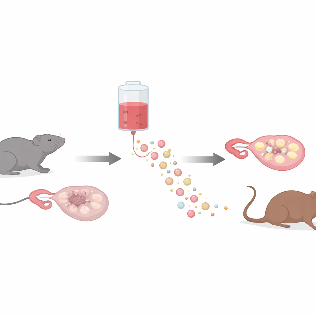
Testing the treatment in a chemotherapy-injured ovary
The team created a rat model of chemotherapy-induced ovarian failure by giving the drug cyclophosphamide, which is known to damage ovarian tissue. After this treatment, the rats showed clear signs of poor health: rough fur, lethargy, disrupted reproductive cycles, and hormone patterns typical of ovarian failure—high levels of stimulating hormones (FSH and LH) and low levels of hormones made by the ovary (estradiol and AMH). The animals were then divided into groups that received either a placebo or one of three doses of MenSCs‑Exos delivered into the bloodstream. Over the next two weeks, the researchers tracked body weight, outward condition, hormone levels, and, after sacrifice, the internal structure of the ovaries and the number of follicles at different stages of development.
How tiny vesicles protected ovarian cells
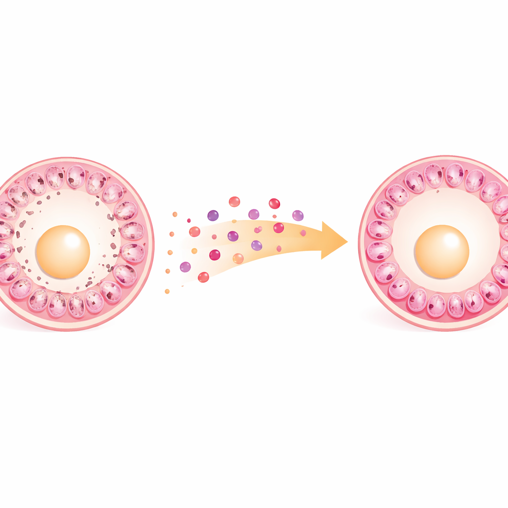
Rats treated with medium or high doses of MenSCs‑Exos regained weight and overall vitality, and their reproductive cycles began to resemble those of healthy controls. Blood tests showed hormone levels moving back toward normal: estradiol and AMH rose, while FSH and LH fell compared with placebo-treated rats. Microscopic examination of the ovaries revealed that MenSCs‑Exos helped preserve and rebuild ovarian tissue. There were more follicles of all stages—primordial, primary, secondary, and antral—and fewer degenerating (atretic) follicles. To probe the underlying mechanism, the researchers focused on two key proteins that control cell death and survival: Bcl‑2, which protects cells, and Bax, which promotes cell suicide. In the damaged ovaries, MenSCs‑Exos increased Bcl‑2, reduced Bax, and raised the Bcl‑2/Bax ratio, all consistent with reduced apoptosis of the granulosa cells that nurture developing eggs.
Finding the sweet spot for dosing
Interestingly, more was not always better. While both the medium and high doses improved health, hormones, and ovarian structure compared with placebo, the highest dose did not offer clear additional benefits over the medium dose. The lowest dose, on the other hand, produced noticeably weaker effects. This pattern suggests there is an optimal therapeutic window—around the medium dose used here—where exosomes provide strong protection and repair without obvious gains from further escalation, an important consideration for designing future clinical therapies.
What this could mean for future fertility care
For now, these findings apply only to rats, and many steps remain before such a treatment could be offered to patients. Still, the work shows that exosomes from menstrual blood–derived stem cells can partially restore hormone balance, ovarian structure, and follicle numbers after chemotherapy, apparently by shielding key ovarian cells from programmed death. Because exosomes are cell-free, scalable, and potentially safer than whole-cell transplants, they may one day become a practical option to help women maintain or regain ovarian function after cancer treatment—provided future studies confirm their safety, ideal dose, and best delivery route in humans.
Citation: Cheng, X., Wu, Y., Cheng, L. et al. Menstrual blood-derived stem cell exosomes improve ovarian function in chemotherapy-Induced POF rats via apoptosis regulation. Sci Rep 16, 13429 (2026). https://doi.org/10.1038/s41598-026-43562-0
Keywords: premature ovarian failure, chemotherapy and fertility, menstrual blood stem cells, exosome therapy, ovarian regeneration