Clear Sky Science · en
Brain tumor segmentation using dual-stream multiscale 3D-UNET with dense net and spatial attention
Why this matters for patients and doctors
Brain tumors are among the most feared cancers, and finding them quickly and outlining them clearly on scans is vital for planning surgery, radiation, and other treatments. This study presents a new computer vision method that can trace the detailed shape of brain tumors on MRI scans with striking accuracy, aiming to support radiologists with faster and more consistent results.
How brain scans reveal hidden trouble
Modern brain imaging uses several types of MRI scans, each highlighting different tissue features. Some emphasize the tumor core, others the surrounding swelling, and others show how fluid changes around the growth. Doctors traditionally draw tumor borders by hand on these scans, a time-consuming process that can vary from one expert to another. Automated methods based on deep learning have improved this task, but many still struggle with tumors that have irregular shapes, blurry edges, or strong differences in brightness from one image to the next.
A smarter way to read MRI pictures
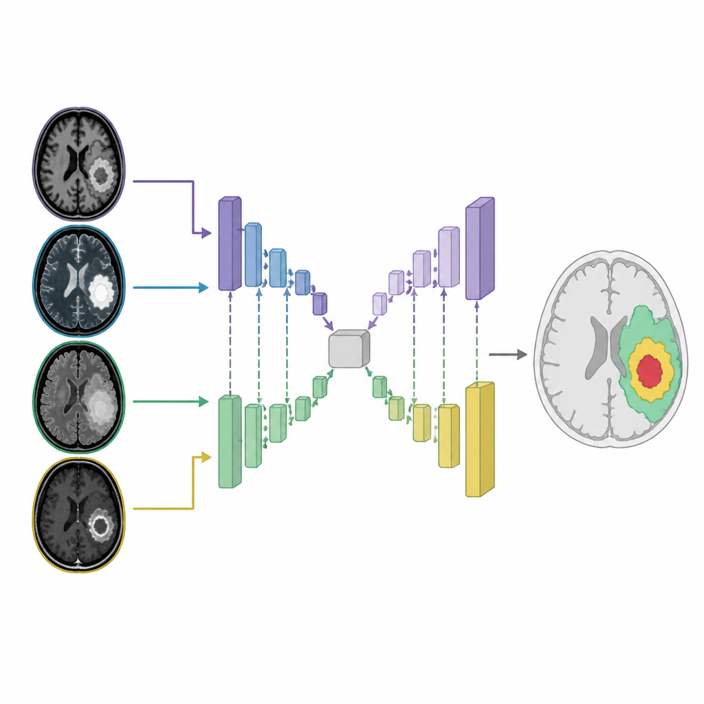
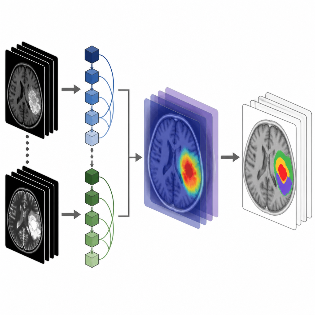
The authors designed a new model that treats the segmentation problem as a 3D task, looking at the brain volume slice by slice instead of flat images alone. Their network has two parallel “streams” that process different MRI inputs at the same time, capturing both fine details and broad context. Inside these streams, a structure called DenseNet passes information forward efficiently so that early details, like tiny edges, are preserved and reused at deeper levels. The model also works at multiple scales, learning from both small features and large regions, which helps when tumors vary greatly in size and shape.
Letting the model focus where it counts
To sharpen its view of problem areas, the network uses a spatial attention mechanism. This component learns to highlight regions of the scans that are more likely to contain tumor tissue, while downplaying normal brain background. Effectively, the model “looks harder” at suspicious areas and “ignores” less informative zones. The researchers further improved learning by carefully preparing the data: they normalized intensities, resized volumes to fit memory limits, and used data augmentation to compensate for classes with fewer examples, such as small enhancing tumors.
How well does it work in practice
The method was trained and tested on the BraTS 2020 brain tumor dataset, a widely used collection of expertly labeled MRI scans, and then checked again on the newer BraTS 2021 set. Across key tumor regions — the active enhancing part, the tumor core, and the full extent including surrounding swelling — the model achieved Dice scores close to 1, indicating almost perfect overlap with expert outlines. It also showed very high sensitivity and specificity, meaning it almost always found tumor tissue when it was present and rarely mislabeled healthy tissue. Importantly, these strong results held up on the separate 2021 dataset, suggesting the approach can generalize beyond the images it was originally trained on.
What this could mean for future care
In plain terms, this study shows that a carefully designed dual-stream, multiscale deep learning model can turn complex MRI data into precise 3D tumor maps that closely match expert work. While the system still needs optimization for speed and broader testing in real clinics, its performance hints at tools that could help radiologists save time, reduce uncertainty, and provide more consistent guidance for surgeons and oncologists when treating people with brain tumors.
Citation: AlSekait, D.M., Zakariah, M., Dubey, P. et al. Brain tumor segmentation using dual-stream multiscale 3D-UNET with dense net and spatial attention. Sci Rep 16, 15416 (2026). https://doi.org/10.1038/s41598-026-43470-3
Keywords: brain tumor segmentation, MRI, deep learning, 3D U-Net, medical imaging