Clear Sky Science · en
Three-dimensional imaging reveals a hierarchical organisation of the myocardial mesh in mammalian hearts
Why the Heart’s Hidden Structure Matters
The strength and reliability of every heartbeat depend not just on how heart cells contract, but on how billions of them are arranged and connected in three dimensions. Yet, despite centuries of study, scientists still argue about the true architecture inside the heart’s muscular wall. This paper uses cutting-edge 3D X-ray imaging to look directly at the fine structure of heart muscle from several mammals, including humans, and reveals that the heart is built as a complex, interwoven mesh rather than simple flat layers or one giant muscle band. Understanding this hidden design helps explain how the heart squeezes efficiently and how its structure may change in disease.
A Closer Look Inside Many Mammalian Hearts
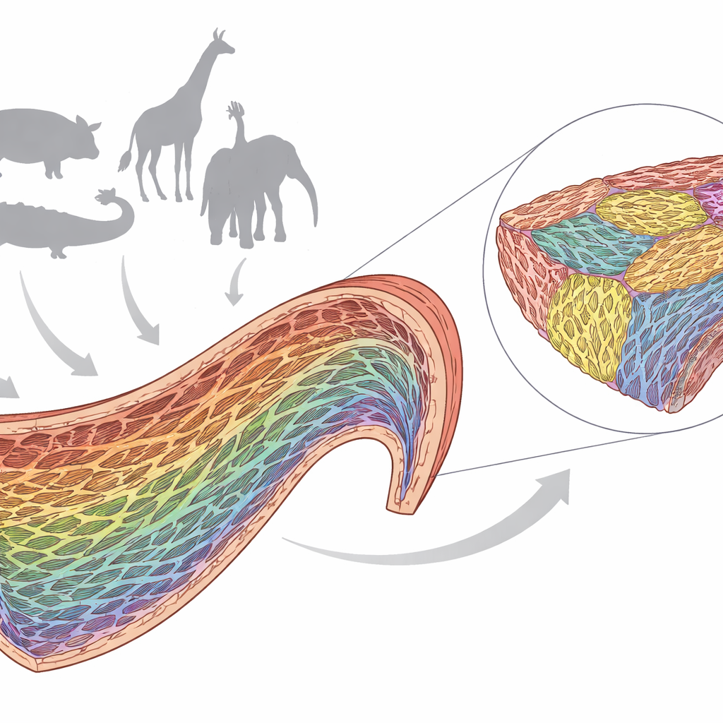
The researchers collected tiny full-thickness samples from the main pumping chamber (left ventricle) of six mammals: human, pig, rabbit, giraffe, elephant, and sei whale. These span a huge range of heart sizes and blood pressures, from small laboratory animals to massive wild species. They soaked the samples in an iodine solution to enhance contrast and then scanned them with high-resolution X-ray microtomography. This technique works like a super-detailed CT scan, allowing the team to reconstruct the tissue in three dimensions with enough clarity to recognize individual heart muscle cells and the connective tissue that binds them together.
A Mesh of Cell Clusters, Not Simple Layers
The 3D images showed that, in all species, heart muscle cells (cardiomyocytes) are arranged as branching chains that together form a continuous three-dimensional mesh. These cells are bundled into small groups held together by fine connective tissue, and these groups are separated from their neighbors by narrow spaces or “clefts” filled with looser tissue, blood vessels, and nerves. Earlier descriptions often called such groupings “sheets” or “sheetlets,” suggesting flat, layered plates. The new reconstructions reveal a far more irregular picture: these units, which the authors simply call “aggregates,” vary widely in thickness, shape, and orientation, even within a single small biopsy. Between species, some hearts show flatter aggregates, while others, like the elephant and whale, display more tube-like structures—but all share the same basic mesh-like organization.
Hidden Hierarchy and Twisting Bundles
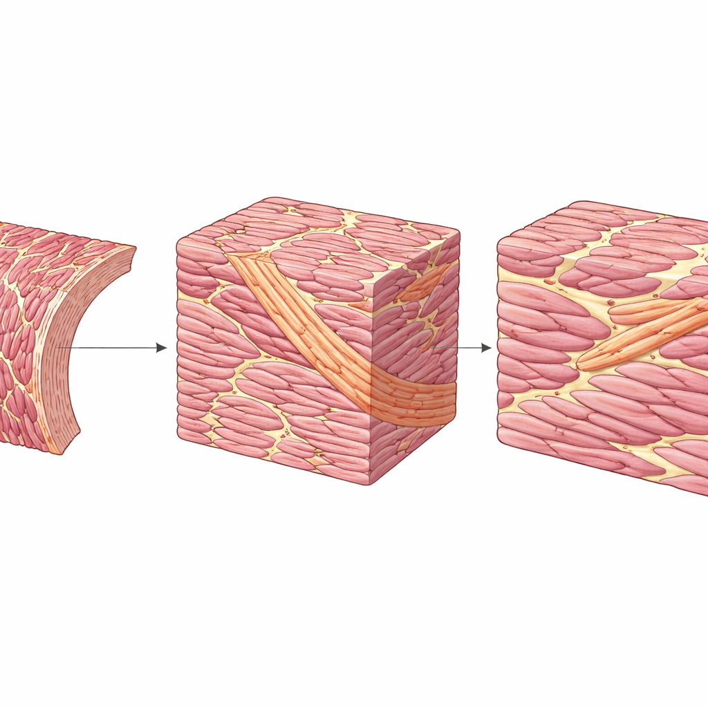
Within this mesh, the team discovered an additional level of organization that had not been clearly described before. Some sets of aggregates are themselves bundled into thicker strands that cut across the surrounding mesh at noticeably different angles. These larger bundles can extend from the outer surface of the heart wall toward the inner surface, twisting and bending as they go. Although they stand out clearly by their orientation, they still branch and reconnect with neighboring tissue rather than forming separate, cord-like structures. Across the wall, the overall direction of the cell chains gradually shifts from one helical angle near the outer surface to the opposite near the inner surface, but locally there are sharp changes in direction of more than 45 degrees between adjacent aggregates. This local variability appears in all the animals studied.
How Microstructure Supports a Beating Heart
These fine-scale arrangements have important functional consequences. Because the heart wall thickens as it contracts, its muscle cannot simply behave like straight, tendon-anchored bundles in skeletal muscle. Instead, the branching mesh and the clefts between aggregates allow small amounts of sliding between neighboring units, helping the wall “repack” itself without tearing. The aggregates and higher-order bundles that tilt away from the main circumferential direction may act as internal counterforces, shaping how the wall thickens and recoils, and helping the ventricle maintain an efficient shape during pumping and filling. This view matches experimental work suggesting that populations of cells pulling in slightly different directions create a balanced, three-dimensional squeeze.
What This Means for Imaging and Heart Health
The findings challenge simplified models that portray the ventricular wall as neatly layered like an onion or as a single, continuous helical muscle band that could be unwrapped in one piece. Instead, the heart’s muscle appears as a hierarchical mesh, from individual cells up through aggregates and larger bundles. This helps interpret clinical imaging methods such as diffusion tensor MRI, which infer fiber directions indirectly and at much lower resolution. The study suggests that the structural units detected by such scans are large enough to be captured reliably, even if individual cells are not seen. As computer models of the heart become more sophisticated, incorporating this mesh-like, heterogeneous architecture rather than idealized layers or a single band should improve simulations of both normal pumping and disease-related remodeling.
A New Picture of the Heart’s Inner Design
In simple terms, this work shows that the heart’s muscle is neither a stack of flat sheets nor one long muscle ribbon, but an intricate, three-dimensional weave of cell groups and bundles. That weave is similar across very different mammals, yet flexible enough to vary in shape and orientation from place to place. By mapping this hidden architecture in detail, the study provides a structural framework for understanding how the heart thickens, twists, and relaxes with every beat—and offers a more realistic blueprint for future imaging, diagnosis, and computer modeling of heart function.
Citation: Stephenson, R.S., Partridge, J., Jarvis, J.C. et al. Three-dimensional imaging reveals a hierarchical organisation of the myocardial mesh in mammalian hearts. Sci Rep 16, 13435 (2026). https://doi.org/10.1038/s41598-026-43337-7
Keywords: cardiac microstructure, myocardial meshwork, heart muscle architecture, X-ray microtomography, diffusion tensor imaging