Clear Sky Science · en
Optimal nano-silica filler concentration to optimize kinetics, rheology and bonding of self-adhesive composites
Why this matters for everyday dental work
When you get a small cavity filled, your dentist often has to go through several careful steps: prepare the tooth, apply a bonding liquid, then place the filling. New "self-adhesive" flowable composites promise to shorten this routine by bonding and filling in a single step. This study asks a deceptively simple question with big practical consequences: how many tiny silica particles should be mixed into these materials so they flow well, cure properly under the dentist’s light, and still grip the tooth firmly?
One-step fillings and the problem of balance
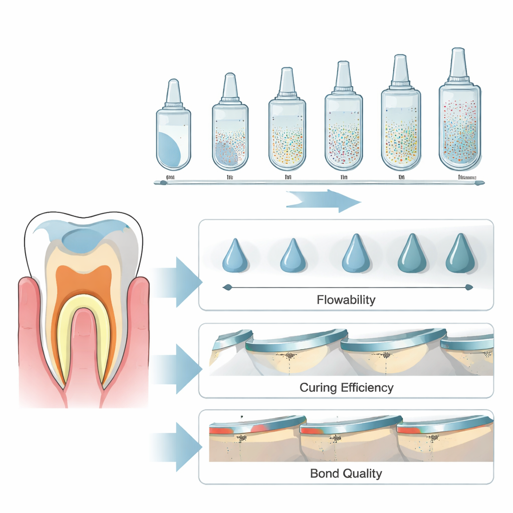
Self-adhesive flowable composites are designed to be low-viscosity pastes that both condition the tooth surface and become the final filling. To work, they must flow into microscopic irregularities in dentin, then harden into a strong, durable solid when exposed to blue curing light. The paste contains a liquid resin and solid fillers, including nano-sized silica particles. Too many particles and the material becomes thick and reluctant to flow; too few and it may shrink more, wear faster, or cure less efficiently. The researchers reframed this as a physics and chemistry puzzle: find the sweet spot in nano-silica content that balances flow, light-driven curing, and bonding to dentin.
How the team tested the new formulations
The team created five experimental versions of a self-adhesive composite that were identical except for the amount of nano-silica, ranging from none to a relatively high fraction. They compared these to a popular commercial product used as a benchmark. With infrared spectroscopy, they tracked how quickly each material’s resin converted from liquid-like molecules into a cross-linked solid under a dental curing light, and how complete that conversion became. A rheometer measured how easily each paste flowed under slow and fast movement, mimicking placement with an instrument versus being squeezed into a tight cavity. To see whether these behaviors translated into real attachment to teeth, they bonded the materials to slices of human dentin, then pulled them apart to measure bond strength and examined the contact zone with special stains and electron microscopy.
What happens when nano-silica goes up and down
The effects of nano-silica were not simple “more is better.” A small addition of particles helped the material react quickly when the curing light was switched on, meaning the network formed faster. However, an intermediate level unexpectedly slowed this early stage. As the nano-silica content continued to rise, the final fraction of resin that converted to solid increased, especially when the light was applied for longer. All versions thinned out when sheared, but their baseline thickness changed in complex ways with particle loading. In practical terms, some pastes flowed into place more easily at low movement, while others only became more workable under higher shear, such as when being spread or pressed by a tool.
Stronger cure did not mean stronger grip
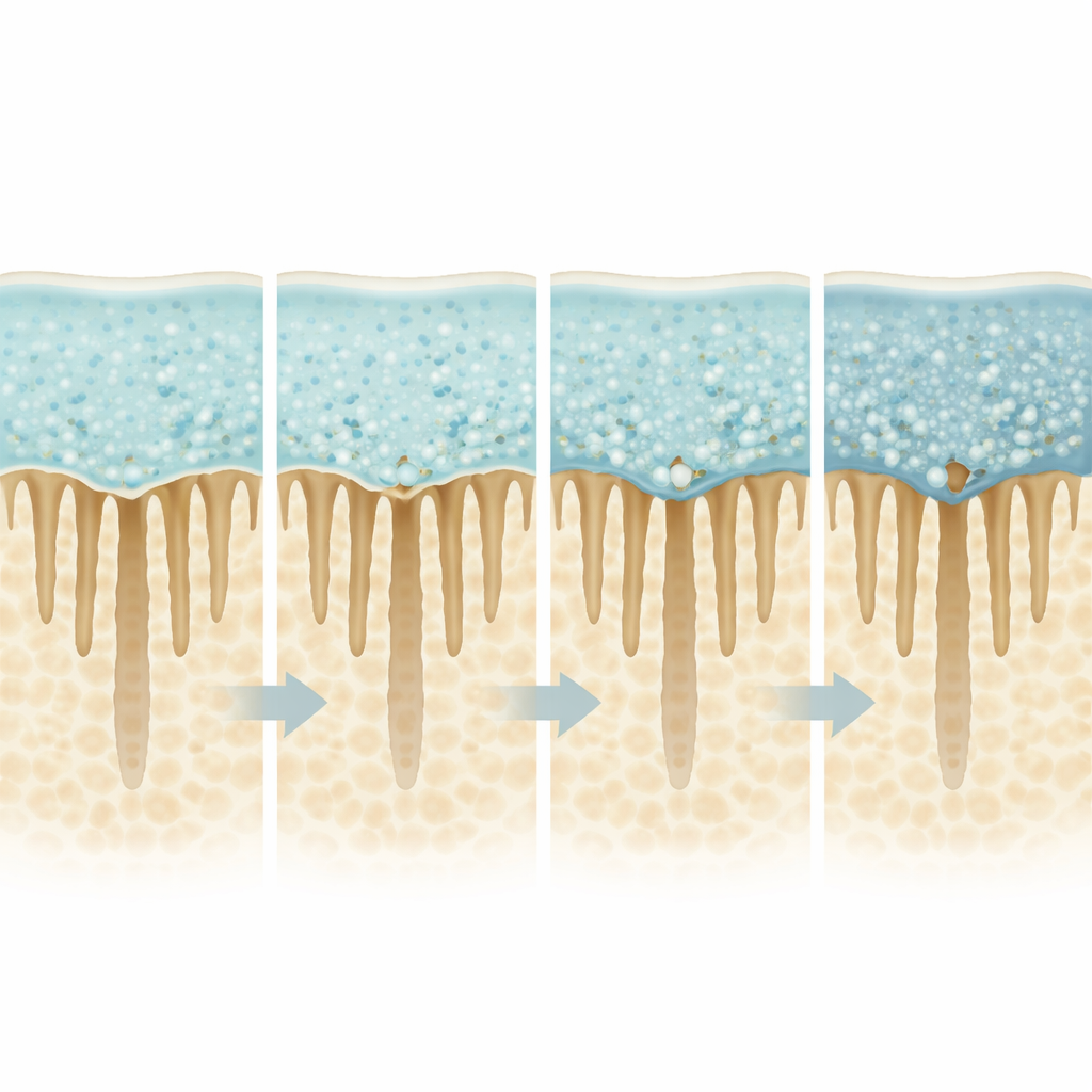
Despite favorable curing behavior at some nano-silica levels, bonding to dentin remained modest. The commercial reference material still held on to the tooth best, and among the experimental versions, the paste without nano-silica performed at least as well as the filled ones. Microscopic images revealed why: instead of forming a thick, interlocked “hybrid” zone with the tooth, all self-adhesive materials mostly sat on top of a smear layer—the thin, ground debris left after drilling. As nano-silica increased, the interface showed more tiny gaps and pores on the composite side. Special staining techniques suggested that collagen fibers in the dentin were often left only partly enveloped by resin, especially at higher particle contents, which leaves the contact zone vulnerable to breakdown over time.
What this means for future fillings
For patients and dentists, the key message is that adding nano-silica to self-adhesive composites changes how fast and how fully they harden, and how they flow, but does not automatically improve their grip on dentin. In fact, very particle-rich versions tended to form more irregular and fragile interfaces. The study suggests there may be a narrow design window where flow, curing, and bonding can be balanced, but current formulations do not yet hit an all-around optimum. To unlock the full promise of truly simple, one-step fillings, future materials will need not only the right amount of nano-silica, but also better control of how the paste wets the tooth surface and how the interface copes with the internal stresses of curing.
Citation: Alves, M., Pereira, P., Silva, D.C. et al. Optimal nano-silica filler concentration to optimize kinetics, rheology and bonding of self-adhesive composites. Sci Rep 16, 12638 (2026). https://doi.org/10.1038/s41598-026-43290-5
Keywords: self-adhesive dental composites, nano-silica fillers, dentin bonding, polymerization kinetics, dental restorative materials