Clear Sky Science · en
ForamJ – A tool for the reproducible, semi-automated analysis of foraminifera micro computed tomography datasets
Why tiny ocean shells matter
On the ocean floor, countless microscopic shells made by single-celled organisms called foraminifera slowly pile up over millions of years. These tiny structures record past seawater conditions, helping scientists reconstruct ancient climates. A new digital tool named ForamJ makes it faster and easier to turn three-dimensional X-ray scans of these shells into hard numbers, opening the door to larger and more detailed climate and ecology studies based on these minute fossils. 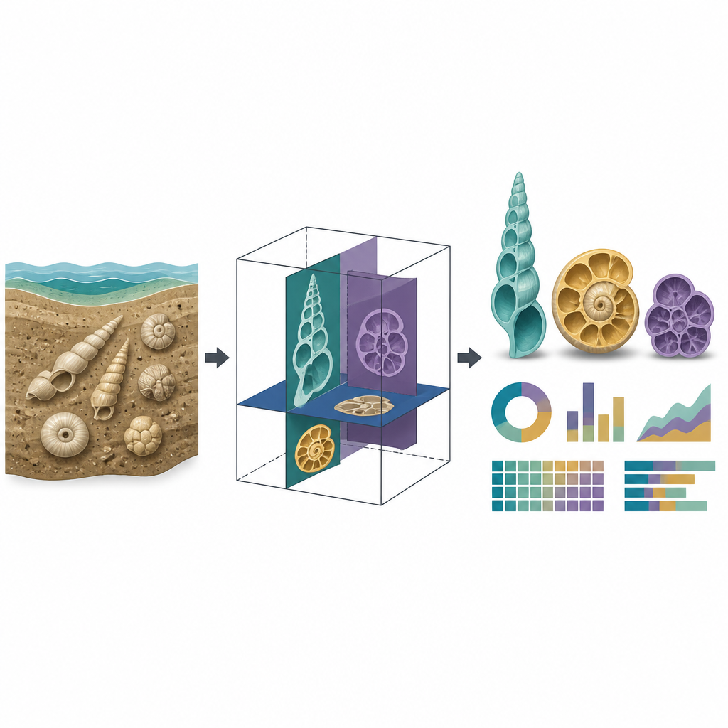
A digital magnifying glass for fossil shells
Micro computed tomography is like a medical CT scan scaled down for millimetre-sized objects. It lets researchers see both the outside surface and the internal chambers of foraminifera without breaking them apart. Until now, scientists wanting to measure features such as shell volume or wall thickness have had to rely on slow, manual tracing in expensive commercial programs. That limited how many specimens could be analysed and made it difficult for different labs to follow exactly the same steps, which is important when building global climate records from many sites.
A free tool built for everyday computers
ForamJ is a free plugin that runs inside the widely used ImageJ and Fiji image software, which already support many kinds of biological imaging. The authors designed it with five practical goals in mind: it should be easy to use for beginners, work on standard laptops, rely only on open software, produce consistent numeric outputs, and handle many scans in one run. ForamJ guides the user through a series of steps, from opening greyscale image stacks to setting the scale, cleaning away sediment packed inside chambers, and separating the shell from the background so that measurements can be taken in three dimensions.
From raw scan to meaningful shell traits
Under the hood, the plugin applies a chain of image operations to each scan. It smooths away noise, keeps only the largest object so that stray grains are removed, and builds a "filled" version of the shell where open spaces are closed so that total volume and surface area can be estimated. It then splits the outer wall from the internal walls between chambers and, using built-in tools, calculates how thick these parts are. ForamJ also isolates the empty space of each chamber and uses a semi-automated method to divide that space into separate chambers even if some walls are partly dissolved. For each chamber it records its volume, position and brightness, and it saves both the processed images and all settings used, so that results can be checked or repeated later. 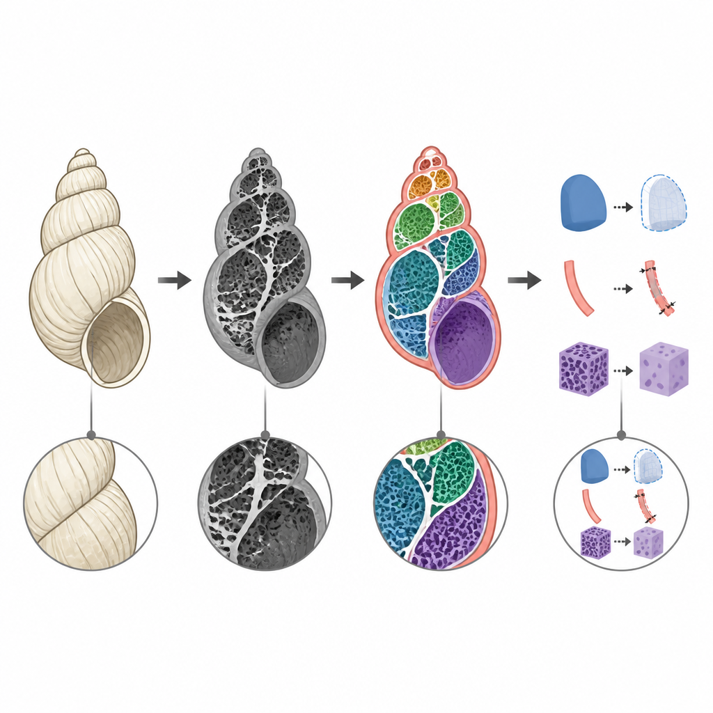
Testing the tool on ancient and modern shells
To demonstrate what ForamJ can do, the researchers applied it to two different sets of foraminifera. First, they examined seven well preserved bottom-dwelling species from 56 million year old sediments drilled off New Jersey. By comparing ForamJ output with painstaking manual work in a commercial program, they found that shell volume differed by about one percent and the match in detailed segmentation was extremely close, while processing time per specimen dropped to a few minutes on an ordinary laptop. The measurements revealed large differences among individuals in shell size, wall thickness, porosity and the way chamber volumes grow from the centre outward.
Exploring the variety of floating shell designs
The team then turned to an open online collection of scans from seven species that float in the upper ocean. ForamJ quickly produced a suite of measurements across all of them, showing a wide range of shell strategies. Some species had very thin walls and extremely porous tests with many chambers, while others built thicker, denser shells with fewer but larger chambers. Growth curves tracing how each new chamber adds volume showed patterns that differed from species to species and even between individuals, pointing to links between shell design, ecology and life cycle that can now be explored more systematically.
What this means for climate and ocean studies
By packaging foraminifera-specific measurements into a simple, shared workflow, ForamJ turns complex three-dimensional scan data into standard numbers that any lab can generate and compare. Because it is free, runs on non-specialist computers, and can process many scans at once, it lowers both cost and time barriers for studies that rely on large sample sets. In practical terms, this means scientists can better track how shell shape, wall thickness and internal structure vary across species, locations and time, improving how we read past ocean conditions from these tiny but information-rich fossils.
Citation: Trend, J., Borges, F.A. & Babila, T.L. ForamJ – A tool for the reproducible, semi-automated analysis of foraminifera micro computed tomography datasets. Sci Rep 16, 14818 (2026). https://doi.org/10.1038/s41598-026-43276-3
Keywords: foraminifera, micro CT, image analysis, paleoceanography, open source software