Clear Sky Science · en
Transformer-enhanced deep ensemble for multi-class liver disease classification using computed tomography images
Why Smarter Liver Scans Matter
Liver disease is quietly becoming a global health crisis, but spotting it early on medical scans can be surprisingly hard, even for experts. This paper explores how modern artificial intelligence can help doctors read routine CT scans more accurately, sorting patients into three common and serious liver problems—fatty liver, cirrhosis, and liver cancer—without extra tests. By combining two powerful AI ideas, convolutional neural networks and transformer attention, the authors build a system that comes close to a highly reliable second opinion for radiologists.
Three Common Liver Problems, One Big Challenge
The liver sits at the center of the body’s chemistry lab, handling metabolism, detoxification, and vital protein production. When it is damaged by long-term fat buildup, scarring, or tumors, the consequences can ripple through nearly every organ system. Fatty liver disease now affects roughly a third of the global population, and cirrhosis and liver cancer account for millions of deaths each year. Yet on CT scans, these conditions often blend into the gray: early fatty changes may look subtle, cirrhotic scarring can be diffuse rather than focal, and tumors can hide among normal tissue. Traditional lab tests help, but they are not specific to individual diseases. Doctors increasingly rely on imaging to decide who needs close monitoring or treatment, but interpretation varies with experience and workload.
Teaching Computers to See in Medical Images
Over the past decade, deep learning has transformed how computers read images. Convolutional neural networks (CNNs) excel at spotting patterns such as edges, textures, and shapes and have already improved detection of many liver conditions. However, classic CNNs mostly focus on local regions and can struggle with diffuse or subtle changes spread across an organ. Transformers, originally designed for language, bring something new: attention. They learn to weigh relationships between distant regions in an image, recognizing global patterns rather than just local patches. The authors of this study set out to blend both strengths—local detail from CNNs and global context from transformers—into a single, practical system for liver CT scans.
Building a Hybrid Team of Neural Networks
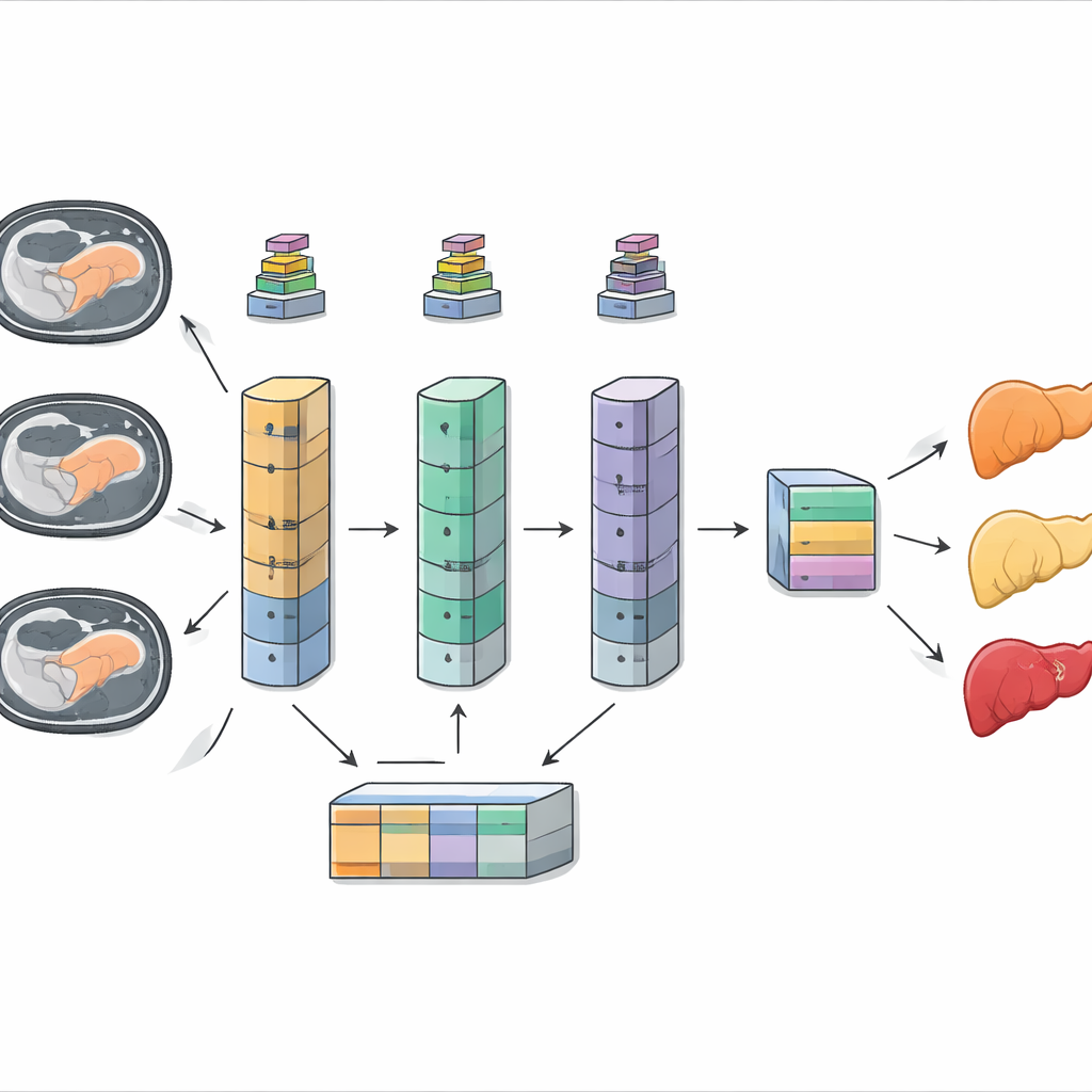
The researchers assembled CT scans from several open datasets, covering 681 patients and over a million individual image slices, representing fatty liver, cirrhosis, and hepatocellular carcinoma (a common form of liver cancer). After standardizing image size and enhancing contrast, they balanced the uneven class distribution with careful data augmentation, slightly shifting, rotating, and zooming the images to mimic real-world variability. Three well-known pretrained CNNs—ResNet50V2, DenseNet121, and MobileNetV2—were first adapted and fine-tuned to classify the three diseases on their own. Each has a different architectural “personality”: ResNet is deep and powerful, DenseNet reuses features efficiently, and MobileNet is lightweight and fast enough for resource-limited settings.
Adding Attention and Fusing Opinions
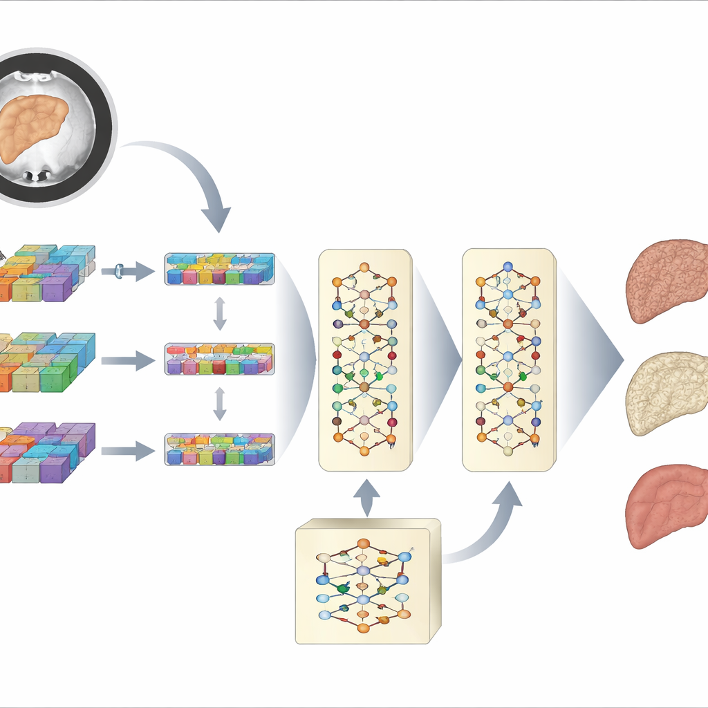
Next, the team extended each CNN with transformer blocks. Instead of stopping at a stack of local features, they reshaped the CNN output into a series of tokens and passed them through multi-head self‑attention layers. These learn which regions of the liver image should “pay attention” to which others, capturing long-range patterns such as widespread scarring or patchy fat deposits. Each hybrid CNN–transformer model produced its own probability for the three disease types, based on all CT slices for a patient rather than single images. Finally, the authors created a hybrid ensemble: they aligned and concatenated the three models’ feature representations and passed them through an additional transformer that learns how to best combine their different viewpoints before making a final decision.
How Well Does the System Work?
The performance gains were striking. On their own, the tuned CNNs reached accuracies between about 69% and 82%, already respectable but with noticeable blind spots—especially for fatty liver and cirrhosis, which often look alike. Adding transformers to each backbone boosted accuracy to 87–93% and greatly improved balance across the three diseases. When all three transformer‑enhanced networks were fused into the ensemble, overall accuracy climbed to 97%, with near-perfect scores for precision, recall, and a robust correlation metric that accounts for class imbalance. Importantly, at the patient level, the ensemble missed no cases of cirrhosis or liver cancer in the test data and showed very few false alarms for fatty liver. Statistical tests confirmed that these gains were not just due to chance but represented a genuine improvement over the best single model.
What This Could Mean for Patients
To a non-specialist, the key message is that this hybrid AI system can turn routine CT scans into a much sharper tool for detecting three major liver diseases at once. By combining different neural networks and giving them an “attention” mechanism, the model learns to notice both fine-grained details and whole-organ patterns that matter for diagnosis. While the approach is computationally heavier than simpler networks and still needs testing across more hospitals and scanners, it points toward practical tools that can sit alongside radiologists, flagging subtle disease, reducing missed cases, and supporting earlier treatment decisions. In short, it suggests a future where smart software helps ensure that no serious liver disease hides in plain sight on a scan.
Citation: Bhardwaj, S., Aggarwal, S., Kumar, N. et al. Transformer-enhanced deep ensemble for multi-class liver disease classification using computed tomography images. Sci Rep 16, 12690 (2026). https://doi.org/10.1038/s41598-026-43256-7
Keywords: liver disease imaging, deep learning diagnosis, CT scan analysis, transformer ensemble, computer-aided radiology