Clear Sky Science · en
Mapping partial agonism of mitragynine at the µ-opioid receptor through molecular dynamics and Markov state modelling analysis
Why a plant compound and pain receptor matter
Opioid painkillers like morphine are powerful but can slow breathing, cause dependence, and be highly addictive. Scientists hope to design new pain relievers that keep the relief while shedding the worst risks. This study focuses on mitragynine, a major ingredient from the Southeast Asian plant kratom, and asks a simple but crucial question: why does it turn the body’s main opioid receptor on only partway, and why does it seem to favor safer signaling routes inside cells?
Two molecules, one pain switch
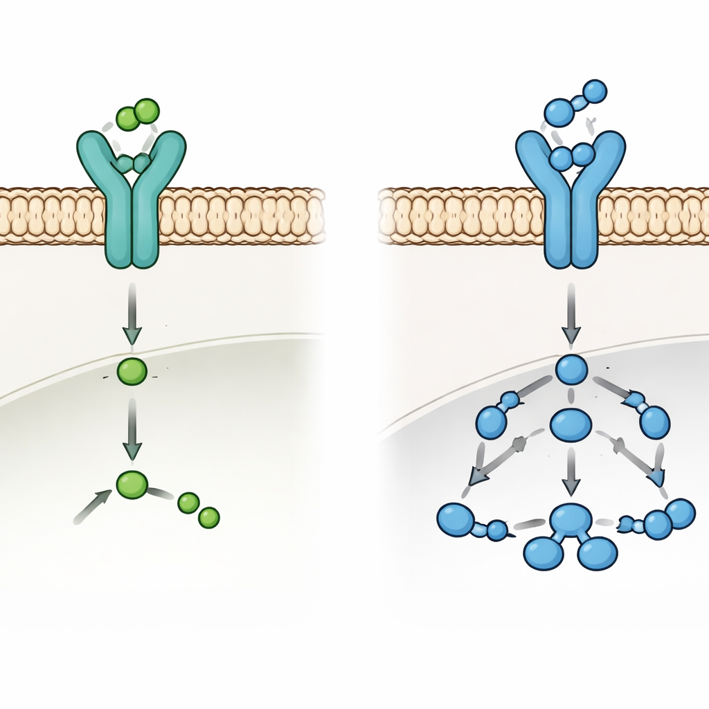
Both morphine and mitragynine act on the same protein on nerve cells, the µ-opioid receptor, which serves as a molecular “dimmer switch” for pain. Morphine is a full agonist: it flips this switch strongly, bringing potent pain relief along with dangerous side effects that are tied to certain signaling pathways. Mitragynine behaves differently. Experiments have shown it gives opioid-like pain relief, but with weaker recruitment of a protein called β-arrestin that is linked to many side effects. In everyday terms, mitragynine appears to press the pain-relief button without fully activating the parts of the system that drive breathing problems and high abuse potential. The current work uses advanced computer simulations to see, at atomic detail, how these two molecules push the same switch into different positions.
Watching the receptor move in virtual time
The researchers built detailed digital models of the µ-opioid receptor sitting in a realistic fatty membrane, once bound to morphine and once bound to mitragynine. They then ran microsecond-scale molecular dynamics simulations, which are essentially physics-based movies showing how every atom jiggles and shifts over time. By tracking how much the receptor’s backbone wandered, how flexible key loops and helices became, and how compact the overall structure stayed, they could infer how stable or restless the receptor was under each drug. Morphine kept the receptor relatively rigid and compact, in line with a firmly “on” position. Mitragynine, by contrast, made certain inner regions—especially those that contact signaling proteins inside the cell—more mobile and variable, hinting that it does not fully lock the receptor into its active shape.
Strong grip, softer push
Interestingly, energy calculations suggested that mitragynine may actually bind more tightly to the receptor than morphine, largely through snug hydrophobic (oil-like) contacts. Yet this strong grip did not translate into stronger activation. Instead, mitragynine produced a more dynamic pattern of hydrogen bonds and subtle shifts in the receptor’s core. To dig deeper, the team turned to Markov state models, a way of piecing together many short structural snapshots into a map of the receptor’s preferred shapes and the routes between them. This analysis showed that morphine funneled the receptor into a deep “active-like” valley on the energy landscape, corresponding to conformations that couple efficiently to downstream partners. Mitragynine spread the receptor’s behavior across a broader set of intermediate shapes, with only limited sampling of fully active conformations.
Lingering in the middle states
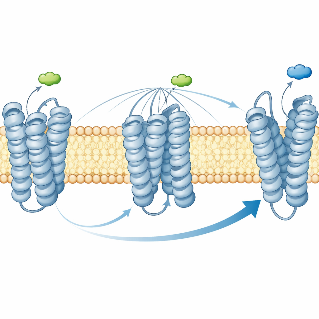
Beyond which shapes are favored, timing matters. The Markov models allowed the authors to estimate how quickly the receptor moves between closed, partly open, and open states. With morphine bound, transitions from intermediate or closed shapes into the open, signaling-ready form happened on the order of hundreds of nanoseconds, reflecting relatively easy access to the “on” state. With mitragynine, those same transitions stretched into microseconds—an order of magnitude slower—meaning the receptor lingered in intermediate states and reached the fully open form only rarely. Overall, the receptor under mitragynine spent much more time in these middle-ground conformations, which are thought to favor G-protein signaling while being less hospitable to β-arrestin binding.
What this means for safer pain relief
Put together, the simulations offer a mechanistic picture of mitragynine as a partial and biased activator of the µ-opioid receptor: it holds on tightly but coaxes the receptor mainly into halfway-on shapes that support G-protein signaling more than β-arrestin engagement. For a layperson, this means mitragynine makes the body’s main opioid switch glow rather than blaze, tilting it toward pain relief with potentially fewer classic opioid harms. While this work is purely computational and does not by itself prove safety in humans, it provides a quantitative framework and structural blueprint that drug designers can use to craft next-generation opioid medicines that follow mitragynine’s lead—strong on pain relief, gentler on side effects.
Citation: Bahari, M.N.A., Azmi, L., Fei, L.C. et al. Mapping partial agonism of mitragynine at the µ-opioid receptor through molecular dynamics and Markov state modelling analysis. Sci Rep 16, 12528 (2026). https://doi.org/10.1038/s41598-026-43251-y
Keywords: µ-opioid receptor, mitragynine, partial agonist, biased signaling, kratom