Clear Sky Science · en
Increased type 3 deiodinase reduces hippocampal T3 signaling and is associated with cognitive impairment in a sporadic Alzheimer’s disease animal model
Why brain hormones matter for memory loss
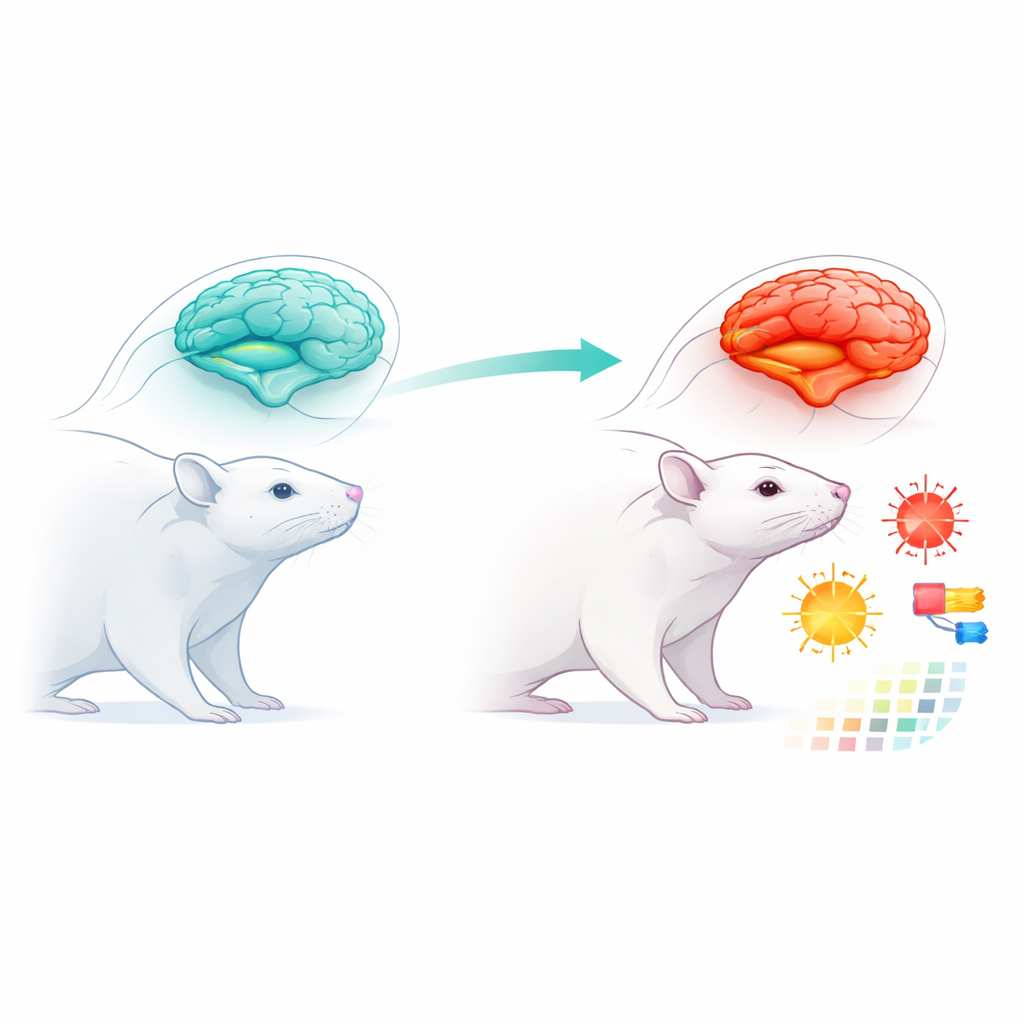
Alzheimer’s disease is usually linked to sticky protein clumps and tangled fibers in the brain, but this study asks a different question: what if part of the problem is that brain cells are running low on a key hormone that keeps them energized and resilient? The researchers focused on a thyroid hormone called T3, which helps brain cells in a memory center called the hippocampus stay healthy. Using a rat model of sporadic Alzheimer’s disease, they explored how changes in local hormone control, inflammation, and chemical stress might work together to damage memory.
A closer look at a memory disease in the lab
To probe this idea, scientists used a well-established method to mimic sporadic Alzheimer’s disease in rats. They injected a compound called streptozotocin (STZ) directly into the brain’s fluid spaces, then followed the animals for 16 weeks. During this time the rats lived under normal conditions, and at the end of the period their memory was tested using a simple task: recognizing a new object compared with a familiar one. Healthy rats naturally spend more time exploring something new, showing they remember what they have seen before. In contrast, the STZ-treated rats failed to distinguish new from old objects, revealing clear problems in both short-term and long-term memory.
Inflammation and chemical stress in the brain
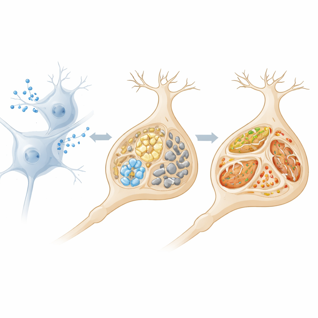
When the researchers examined the animals’ brains, they found that the Alzheimer-like rats showed strong signs of inflammation. Levels of two key inflammatory messengers, IL-6 and TNF-α, were much higher than in control animals. At the same time, brain tissue showed increased protein damage linked to oxidative stress—a kind of chemical wear-and-tear caused by reactive molecules. The main antioxidant guardian, glutathione, and other protective sulfhydryl groups were depleted. Although certain antioxidant enzymes became more active, this boost looked more like a strained, last-ditch response than an effective defense, suggesting that the brain’s protection system was being overwhelmed.
When local hormone brakes go into overdrive
The core of the study centers on how the brain handles thyroid hormones locally. Rather than relying only on hormone levels in the blood, the brain uses special enzymes to fine-tune how much active T3 each cell sees. One of these enzymes, type 3 deiodinase (D3), acts like a brake: it inactivates T3 and its precursor, reducing their availability inside cells. In the Alzheimer-like rats, both the gene and protein levels of D3 in the hippocampus were strongly increased. At the same time, genes that normally respond to T3 were turned down, signaling that brain cells were experiencing a state of “local low thyroid” even though the rest of the body might appear normal. The authors link this shift to the ongoing inflammation and oxidative stress, which are known to push D3 into overactivity.
Energy factories under strain
This apparent T3 shortage did not happen in isolation. The team found that several markers of mitochondrial health—molecules involved in building and controlling the cell’s energy factories—were reduced in the STZ-treated rats. Proteins that support energy production and help prevent cell death dropped, while stress signals from the cell’s protein-folding machinery, the endoplasmic reticulum, rose sharply. Together, these changes paint a picture of neurons caught in a vicious cycle: inflammation and chemical stress boost D3, D3 strips away active thyroid hormone, and the resulting hormone deficit weakens mitochondria and protein handling, making brain cells even more vulnerable to damage and death.
What this means for understanding and treating Alzheimer’s
For a general reader, the key message is that Alzheimer’s disease may not only be about protein plaques and tangles. This work suggests that in at least one widely used animal model, memory loss goes hand in hand with a local hormone imbalance in the hippocampus, driven by inflammation and oxidative stress. By cranking up the activity of D3, the brain effectively starves its own cells of T3, undermining energy production and survival pathways. The authors argue that this “hidden” thyroid problem inside the brain may be an important amplifier of neurodegeneration. If similar mechanisms operate in people, targeting D3, restoring T3 action locally in the brain, or easing the redox and inflammatory burden could open new, more precise treatment avenues for slowing or preventing cognitive decline.
Citation: Marschner, R.A., Ribeiro, R.T., Gayger-Dias, V. et al. Increased type 3 deiodinase reduces hippocampal T3 signaling and is associated with cognitive impairment in a sporadic Alzheimer’s disease animal model. Sci Rep 16, 13666 (2026). https://doi.org/10.1038/s41598-026-43232-1
Keywords: Alzheimer’s disease, thyroid hormone, hippocampus, oxidative stress, neuroinflammation