Clear Sky Science · en
Highly sensitive electrochemical detection and quantification of opium derived morphine sulfate using cysteamine loaded MWCNTs@V2O5 telluride composite
Why This Matters for Pain Medicine and Safety
Powerful painkillers like morphine can be life-changing for people with cancer or severe injuries, but they also carry risks of addiction, overdose, and illegal use. Doctors and forensic specialists need quick, accurate ways to measure how much morphine is really in a person’s body fluids. This study describes a new tiny electronic sensor that can detect very low levels of morphine sulfate in blood serum, using advanced nanomaterials to make the test faster, more sensitive, and potentially easier to use outside of large, specialized laboratories.
Turning a Painkiller into a Measurable Signal
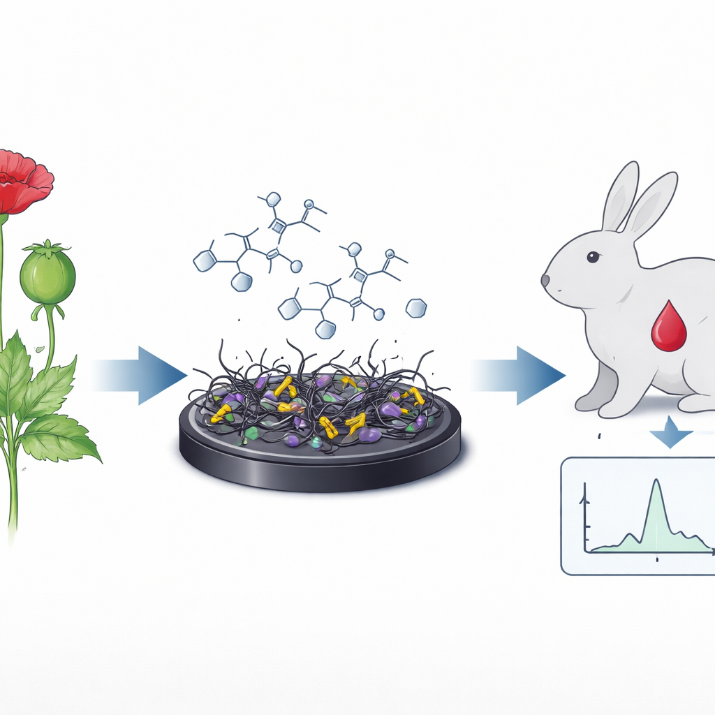
Morphine comes from the opium poppy, a plant long known for easing pain but also for causing dependence. In hospitals, morphine sulfate is one of the strongest drugs used to control severe and chronic pain, especially in cancer patients. Yet the same properties that make it effective also mean that the dose has to be carefully controlled. Traditional laboratory methods for measuring morphine in blood or urine—such as gas or liquid chromatography—are precise but slow, expensive, and require bulky equipment. The authors set out to build an electrochemical sensor: a small, modified electrode that turns the presence of morphine molecules into a measurable electrical current.
Building a Tiny High-Tech Surface
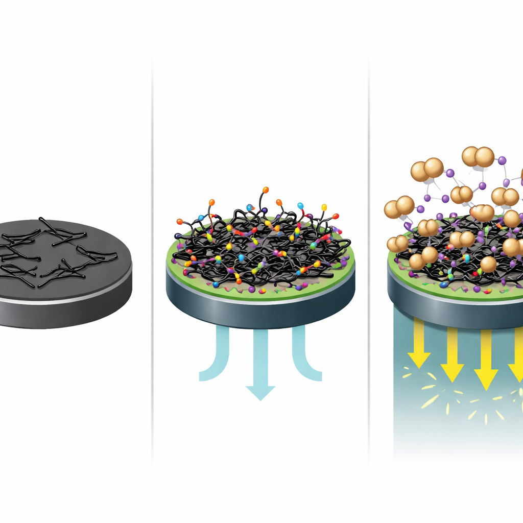
To create this sensor, the researchers engineered a layered nanomaterial on top of a standard glassy carbon electrode. The base building blocks are multi-walled carbon nanotubes—microscopic hollow tubes of carbon that conduct electricity very well and create a large surface area for chemical reactions. These nanotubes were first oxidized to add reactive sites, then coated with vanadium pentoxide and telluride, two conductive materials that further improve electrical performance and increase the number of active spots where reactions can occur. The result is a composite called MWCNTs@V2O5/Te, which forms nanorod-like structures with a rough, porous surface ideal for sensing.
Making the Sensor Seek Out Morphine
A key challenge is getting morphine molecules in a complex liquid like blood serum to selectively attach to the electrode surface. To solve this, the team used a small linker molecule called cysteamine. One end of cysteamine binds strongly to the nanocomposite via a sulfur-based bond, while the other end can interact with chemical groups on morphine sulfate, helped by iron provided as ferric cyanide. This creates a kind of molecular “Velcro” that draws morphine close to the electrode. When a voltage is applied, morphine undergoes oxidation and reduction at the surface, and the resulting flow of electrons shows up as peaks in standard electrochemical tests such as cyclic voltammetry and differential pulse voltammetry.
Testing Performance in the Lab and in Blood
The researchers carefully characterized their nanocomposite with electron microscopy, X-ray diffraction, infrared spectroscopy, and UV–visible spectroscopy to confirm its structure and composition. Electrochemical measurements showed that the modified surface had a much larger electrochemically active area and lower resistance to charge transfer than a bare electrode, meaning electrons move more easily during sensing. When exposed to increasing concentrations of morphine sulfate in buffer, the sensor produced steadily growing current signals, with an excellent linear relationship between current and concentration from 10 to 60 micromoles and a very low detection limit of about 0.01 micromoles. The sensor’s response depended on acidity, working best near physiological pH (7.4), and it remained stable over prolonged testing and in the presence of common interfering substances such as glucose and protein.
From Rabbits to Real-World Use
To see how the sensor behaves in realistic conditions, the authors tested it on rabbit serum after injecting morphine sulfate. Blood samples taken at different times showed strong signals when morphine levels were highest and decreasing currents as the drug was cleared from the body, matching expectations about its half-life. In these real samples, the detection limits remained very low, and recovery tests showed that the sensor could accurately measure known added amounts of morphine. Compared with other modern electrochemical morphine sensors reported in the literature, this design offers competitive or better sensitivity along with good selectivity, reproducibility, and stability.
What This Could Mean for Patients and Investigators
Overall, the study demonstrates that a thoughtfully engineered nanomaterial surface can turn a simple electrode into a highly sensitive detector for morphine sulfate in blood. By combining carbon nanotubes, metal oxides, telluride, and a smart linker molecule, the authors created a platform that can pick up very low drug levels, distinguish morphine from common background substances, and operate reliably over time. With further development and packaging, such sensors could help clinicians adjust pain treatment more precisely, assist toxicologists and forensic scientists in confirming narcotics exposure, and support monitoring in settings where fast, on-site results are crucial.
Citation: Shaheen, S., Fatima, B., Hussain, D. et al. Highly sensitive electrochemical detection and quantification of opium derived morphine sulfate using cysteamine loaded MWCNTs@V2O5 telluride composite. Sci Rep 16, 13558 (2026). https://doi.org/10.1038/s41598-026-43216-1
Keywords: morphine monitoring, electrochemical sensor, carbon nanotubes, nanocomposite biosensor, drug detection in serum