Clear Sky Science · en
Artificial intelligence assistance improves endoscopist accuracy for gastric cancer dysplasia and intestinal metaplasia
Why smarter stomach checks matter
Stomach cancer remains one of the world’s deadliest cancers, yet many dangerous changes in the stomach lining are subtle and easy to miss, even for trained doctors using modern cameras. This study asks a simple question with big consequences: can an artificial intelligence (AI) assistant, watching endoscopy videos alongside the doctor, help catch more trouble spots earlier and more reliably? By testing a sophisticated AI system with real patient images and videos, the researchers show that digital help can significantly boost the accuracy of less-experienced endoscopists, especially for the hardest-to-judge changes that often precede cancer.
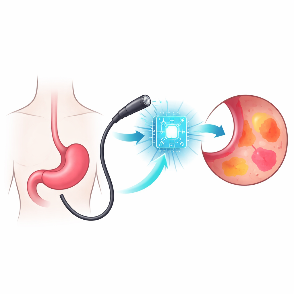
From routine camera exam to data-rich problem
To look for stomach disease, doctors pass a flexible camera, or endoscope, through the mouth into the stomach. They must judge, in real time, whether each patch of tissue is normal, inflamed, precancerous, or cancerous. Some findings, like advanced tumors, stand out clearly. Others—such as dysplasia (early abnormal growth) and intestinal metaplasia (a high‑risk change in the stomach lining)—appear as faint color shifts or delicate texture changes that vary from person to person. In everyday practice, these subtle patterns are easy to overlook, and detection rates differ widely among doctors, which means some patients with high‑risk lesions may leave the exam room undiagnosed.
How the AI co-pilot was built and tested
The team developed an AI system called Venotics‑G using a modern “vision transformer” architecture, designed to scan an image as a whole and notice long‑range patterns. They trained it on over 16,000 endoscopic images from a major hospital, teaching it to recognize several tasks at once: where the scope is in the stomach, whether any abnormal area is present, what type of lesion it might be, and whether high‑risk intestinal metaplasia is visible. For the study, they assembled 1,584 real endoscopy cases from two hospitals. Each case contributed both a single clear still image and a short five‑second video clip in which the suspicious area was well seen. The cases covered stomach cancer, dysplasia, a broad set of benign conditions, high‑risk intestinal metaplasia, and normal or simple gastritis.
Putting AI and human judgment head to head
Six in‑training endoscopists, each with less than three years of experience, reviewed all the cases twice: first relying only on their own judgment, and later, after a one‑week break and with the case order shuffled, reviewing them with the AI’s guidance. The AI alone proved highly accurate on both still images and videos, correctly classifying more than 9 out of 10 focal lesions and high‑risk intestinal metaplasia. More importantly, when the doctors had access to the AI’s suggestions and visual highlight maps, their own performance climbed sharply. Overall accuracy for focal lesions rose from about 75% to nearly 87% for both images and videos, and their combined measure of true‑positive and true‑negative performance (summarized as the area under the ROC curve) improved markedly. Gains were especially large for dysplasia and intestinal metaplasia—precisely the categories that had shown the most variability and lowest baseline accuracy.
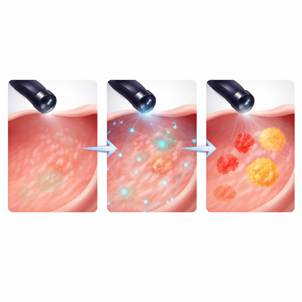
What changed inside the decision-making process
Closer analysis revealed that AI support helped in slightly different ways depending on the condition and whether the doctors were reading images or videos. For dysplasia, the main benefit was nudging readers to recognize more true lesions, while still keeping false alarms low. For intestinal metaplasia, which often appears as diffuse, patchy changes, AI mainly reduced false positives on still images—making the doctors more confident when calling a region normal. In the video setting, the AI could use multiple frames over time, improving both its ability to rule out normal tissue and to flag truly abnormal, widespread patterns. The system’s rapid processing, on the order of milliseconds per frame, means it can keep up with a live endoscopic exam, overlaying attention maps in near real time. At the same time, the authors note that extra on‑screen cues can add cognitive load, and some readers tended to over‑interpret benign inflammation, hinting that training on how to use AI responsibly will be as important as the algorithms themselves.
What this could mean for patients and clinics
Overall, the study shows that an AI assistant can act as an equalizer for less‑experienced endoscopists, making their diagnoses more consistent and more often correct across many types of stomach lesions. Rather than trading fewer misses for more false alarms, the AI tended to improve both sensitivity and specificity together, a sign of true added value rather than mere bias. The work is still an early, carefully curated test, so its impact on day‑to‑day workflows, costs, and long‑term patient outcomes remains to be proven in larger, live clinical trials. But these results support a future in which endoscopy is routinely performed with an AI partner, helping doctors spot high‑risk changes earlier and more reliably, and giving patients a better chance at timely treatment and improved survival.
Citation: Lee, Y.H., Park, G., Kim, J.Y. et al. Artificial intelligence assistance improves endoscopist accuracy for gastric cancer dysplasia and intestinal metaplasia. Sci Rep 16, 12428 (2026). https://doi.org/10.1038/s41598-026-43149-9
Keywords: gastric cancer screening, endoscopy AI, intestinal metaplasia, dysplasia detection, medical image analysis