Clear Sky Science · en
Dynamic wavelet transform normalized multi scale dense attention UNet with Binary Horse Herd Optimization for osteosarcoma diagnosis
Sharper Views of a Dangerous Bone Cancer
Osteosarcoma is an aggressive bone cancer that often strikes children and young adults. Doctors rely on microscope images of bone tissue to decide how much of a tumor is still alive after treatment, a judgment that guides surgery and chemotherapy. But these images are complex, subtle, and time‑consuming to read by eye. This study introduces an artificial intelligence (AI) system that aims to support pathologists by finding and labeling cancer regions in bone tissue slides with remarkable precision.
Why Reading Bone Slides Is So Hard
Under the microscope, osteosarcoma does not look like a neat, solid lump. Instead, islands of living tumor cells, dead tissue, and normal bone weave together in intricate patterns. Traditional imaging and manual review can miss tiny islands of tumor or blur the boundary between diseased and healthy regions. Different experts may disagree on where tumor stops and normal bone begins. These uncertainties can affect biopsy guidance, how much bone is removed in surgery, and how treatment response is judged. The authors argue that smarter computer tools could make these decisions more consistent and less dependent on individual judgment.
Building an End-to-End Digital Assistant
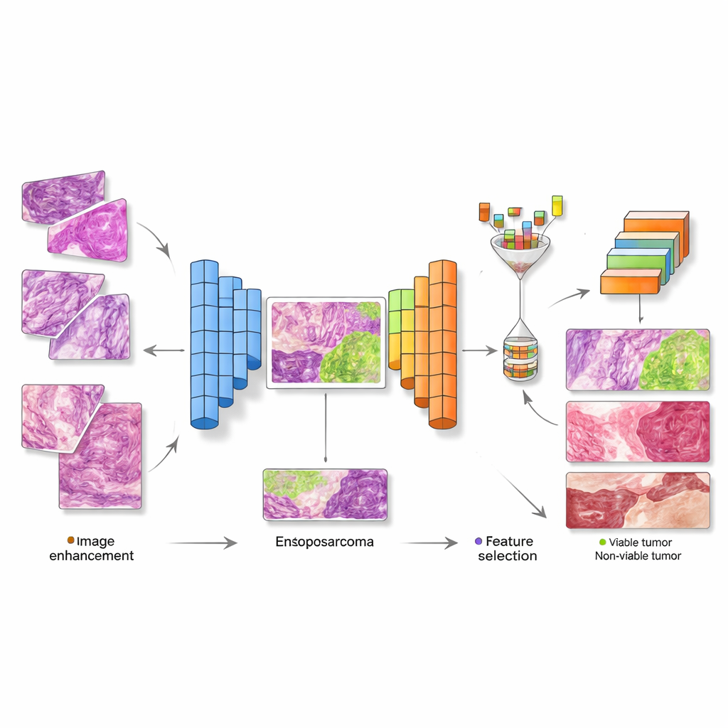
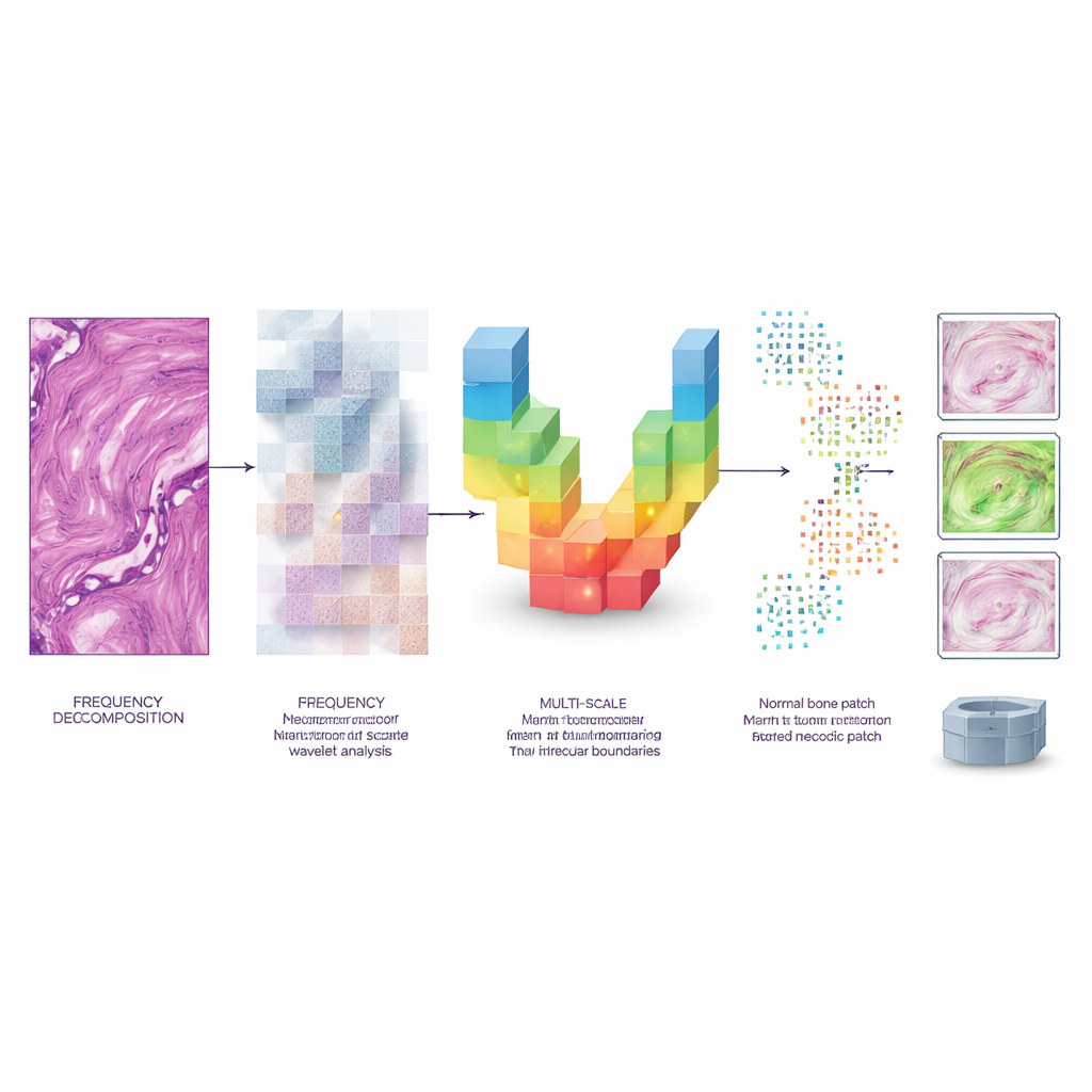
The research team designed a complete, step‑by‑step pipeline that takes in digitized microscope images and produces both a detailed map of tumor regions and a final class label for each image. First, a preprocessing stage uses a mathematical technique called a wavelet transform to clean up noise and boost contrast without washing out fine detail. Next, a specially crafted neural network, shaped like a U and packed with “attention” modules, learns to draw precise borders around abnormal tissue at several scales—from tiny cell clusters to larger structural patterns. This network outputs segmentation masks that show which pixels belong to viable tumor, non‑viable (dead) tumor, or healthy tissue.
Picking Only the Most Telling Clues
Modern deep networks can produce thousands of measurements from each image, but many are repetitive or unhelpful. To avoid confusion and overfitting, the authors add a feature‑selection stage inspired by herd behavior. In this step, a “Binary Horse Herd Optimization” algorithm searches through the large pool of features and keeps only those combinations that best separate tissue types, discarding the rest. This pruning reduces the data size by about 70 percent while preserving the patterns that matter most for diagnosis. The streamlined feature set then feeds into a compact transformer‑based classifier that is tuned by another optimization method nicknamed the Puma Optimizer.
How Well Does It Work?
The system was tested on 1,144 high‑resolution histopathology images from children treated for osteosarcoma at a single medical center over two decades. Each image was labeled as non‑tumor, viable tumor, or non‑viable tumor. Using fivefold cross‑validation, the segmentation network achieved overlap scores (Dice coefficients) above 99 percent and very low boundary errors, outperforming several well‑known medical imaging networks. The final classifier reached about 99.5 percent accuracy, with equally high precision, recall, and specificity across all three classes. In practical terms, this means very few missed tumors and very few healthy regions wrongly flagged as cancer, yet the system still ran fast enough—about 63 milliseconds per image—to be usable in a clinical workflow.
What This Could Mean for Patients
For patients, the promise of this work is not a robot doctor but a more reliable second set of eyes. By sharpening images, tracing tumor contours at multiple scales, and focusing on the most informative patterns, the framework offers pathologists a highly consistent, data‑driven view of which parts of a bone sample are alive tumor, dead tumor, or normal tissue. This could improve how biopsies are targeted, how treatment response is quantified, and how surgeons plan limb‑saving operations. The authors note that more testing on larger, multi‑center datasets is still needed, but their results suggest that carefully designed AI pipelines can bring both accuracy and practical speed to one of the toughest diagnostic tasks in bone cancer.
Citation: Sumathi, S., Maheswari, R., Babu, A.S. et al. Dynamic wavelet transform normalized multi scale dense attention UNet with Binary Horse Herd Optimization for osteosarcoma diagnosis. Sci Rep 16, 12987 (2026). https://doi.org/10.1038/s41598-026-43015-8
Keywords: osteosarcoma, bone cancer imaging, histopathology AI, tumor segmentation, deep learning diagnosis