Clear Sky Science · en
Artificial intelligence based assessment of treatment response in wet age related macular degeneration using paired OCT angiography
Why this matters for sight
Age related macular degeneration is a leading cause of vision loss in older adults, and many people receive repeated eye injections to keep the disease in check. Doctors rely on eye scans to decide whether treatment is working, but reading these scans is difficult and can vary from one specialist to another. This study explores how artificial intelligence can help judge treatment success more consistently, using a newer type of eye scan that maps tiny blood vessels in the back of the eye.
Seeing blood flow without dye
For years, specialists have used dye based imaging to find abnormal blood vessels that threaten central vision. More recently, optical coherence tomography and its offshoot, OCT angiography, have allowed doctors to see fine layers of the retina and its blood flow without a needle stick. These scans can show how abnormal vessels grow, shrink, or change shape during treatment, but the patterns are often subtle and do not always match obvious signs of active disease such as fluid or bleeding. As a result, judging whether a course of injections truly helped, did nothing, or allowed the disease to worsen can be time consuming and subjective.
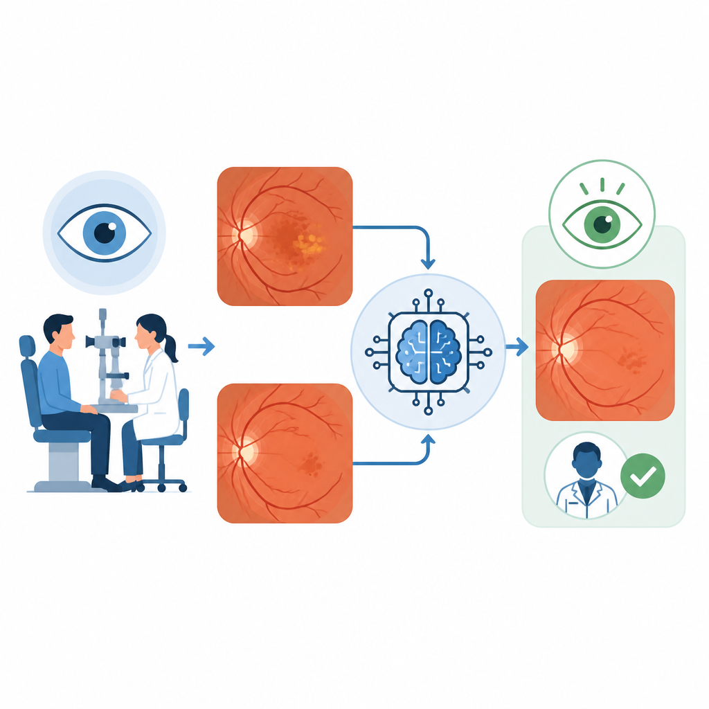
Training a digital second opinion
The researchers gathered more than one thousand pairs of OCT angiography images from people with the wet form of macular degeneration. Each pair showed the same eye before and after a series of anti VEGF injections, a standard drug therapy that slows abnormal vessel growth. Separate cross sectional scans and changes in vision were used to decide whether each treatment course had clearly improved the eye, left it essentially unchanged, or allowed it to worsen. These expert labels served as the reference standard for teaching a deep learning system to sort new image pairs into the same three groups.
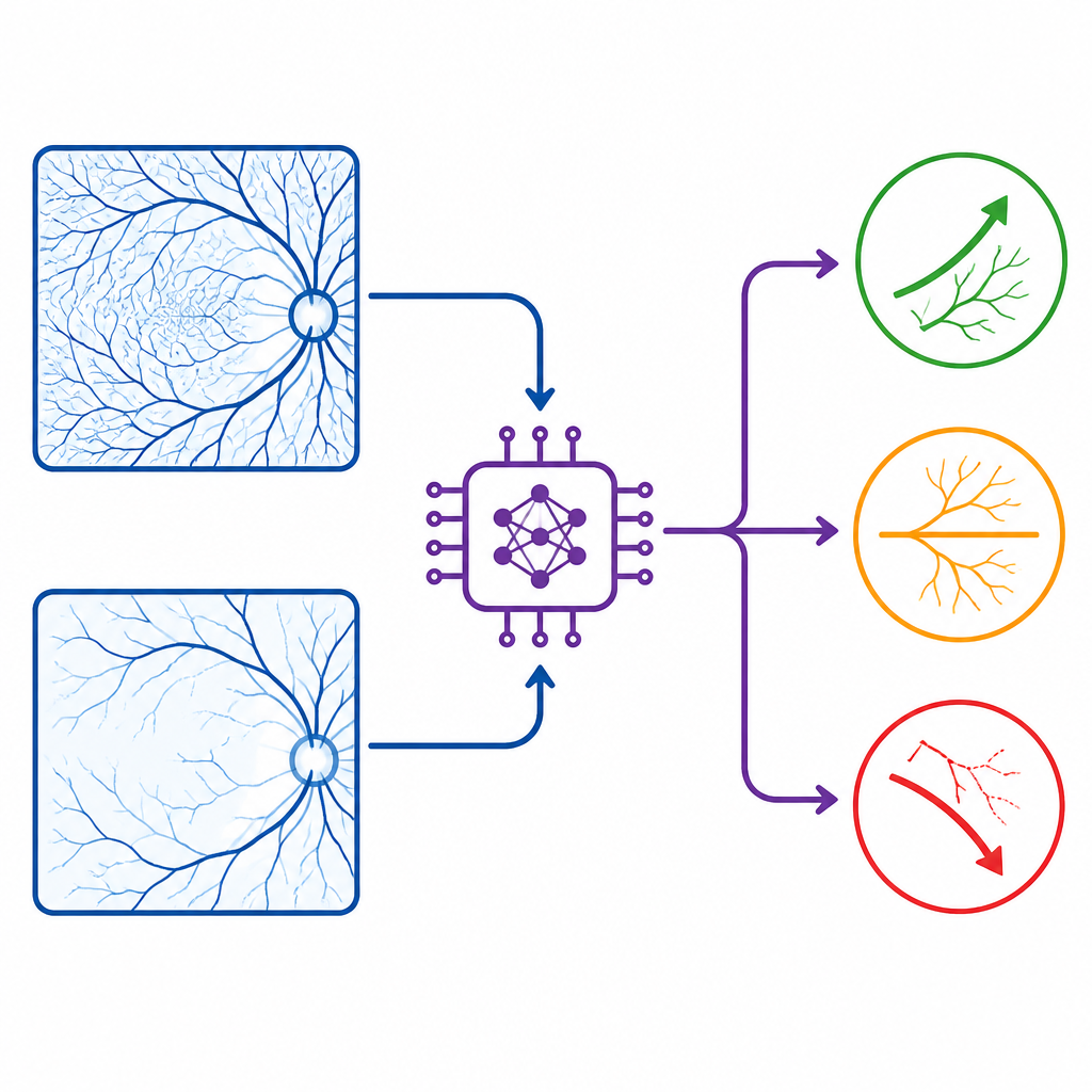
How the computer reads the scans
The team built a model with two parallel branches, one for the pre treatment scan and one for the post treatment scan. For each time point, the system examined several layers of the retina and choroid captured as flat, top down views. It first learned to extract complex visual features from each scan, such as vessel density and branching patterns, then combined information from both time points to produce a single judgment of improved, unchanged, or worsened response. The images were randomly split so that some pairs were used to train the model, some to fine tune it, and a separate set was held back for final testing.
Comparing the computer with experts
On the independent test set, the artificial intelligence system correctly classified treatment response in about eight out of ten image pairs, with especially strong performance in spotting improvements. Two experienced retina specialists reviewed the same image pairs without access to other clinical data. Their combined accuracy was closer to six in ten, and they struggled most with cases where the disease was stable rather than clearly better or worse. Statistical analysis showed that human graders were nearly three times more likely than the model to misjudge whether a treatment course helped, did nothing, or failed, highlighting how challenging these scans can be to interpret by eye alone.
What this could mean for patients
The study suggests that a carefully trained artificial intelligence system can act as a consistent reader of paired vessel scans, offering an objective view of how the abnormal vessels in macular degeneration respond to treatment. While such tools will not replace doctors, they could serve as a decision aid, flagging subtle changes, reducing disagreement between experts, and helping to tailor injection schedules more precisely. In the long term, this approach may support more personalized care and better use of new and existing drugs to preserve vision in people living with this common eye disease.
Citation: Morsy, M.S., Dutta, N.A., Eldessouky, E.I. et al. Artificial intelligence based assessment of treatment response in wet age related macular degeneration using paired OCT angiography. Sci Rep 16, 15405 (2026). https://doi.org/10.1038/s41598-026-42999-7
Keywords: age related macular degeneration, optical coherence tomography angiography, artificial intelligence, treatment response, retinal imaging