Clear Sky Science · en
KIF20A promotes cervical cancer progression by interacting with CLIP1
Why this research matters to women’s health
Cervical cancer remains one of the leading causes of cancer deaths in women worldwide, especially in countries where screening and vaccination are still hard to access. While we know that persistent infection with certain human papillomaviruses is the main trigger, doctors and scientists are still working to understand why some infected cells turn into aggressive tumors and others do not. This study zooms in on two tiny cellular machines, called KIF20A and CLIP1, and shows how their partnership helps cervical cancer cells grow and spread—pointing to a new way to catch and possibly treat the disease.
Looking for new warning signs inside tumor cells
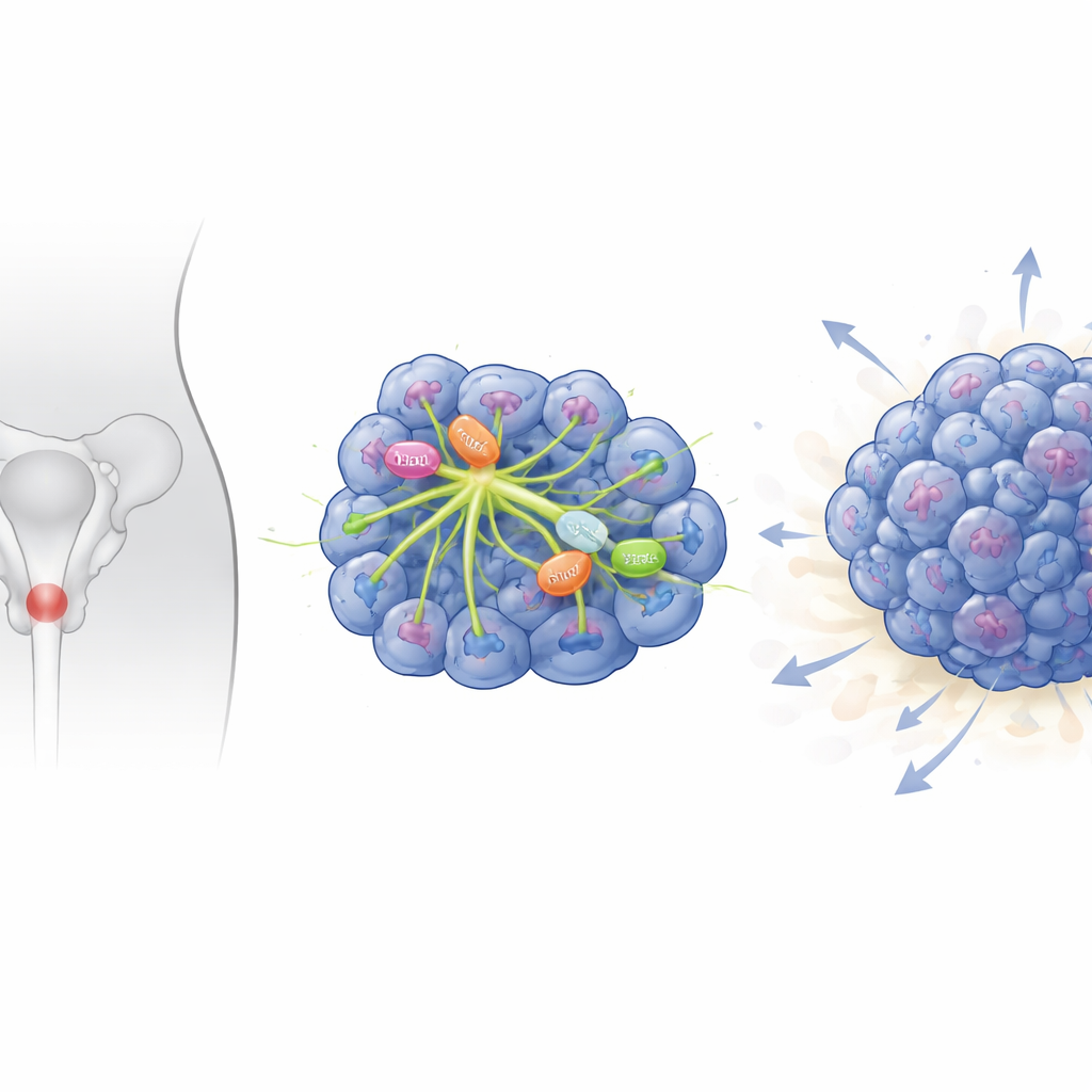
The researchers began by searching large public databases that contain genetic information from hundreds of cervical cancer patients and healthy controls. They compared which genes were turned up or down in tumor tissue versus normal cervical tissue across three independent datasets. Out of thousands of genes that changed, 426 were consistently more active in cancer. Many of these genes were involved in how cells divide and copy their DNA—processes that go awry in cancer. Among them, a small group of “motor” genes that move along the cell’s inner scaffolding stood out, and one in particular, called KIF20A, showed especially strong increases in cervical cancer samples.
A motor protein that is overactive in cervical tumors
To see if this pattern was real in patients, the team examined cervical tumor tissues collected from the hospital and compared them with nearby non-cancerous tissue from the same women. They measured both the genetic message (mRNA) and the actual protein made from the KIF20A gene. In more than 300 tumor samples, KIF20A levels were much higher than in 22 normal tissues, and this was true in the major types of cervical cancer. Staining thin slices of tissue under the microscope showed that cancer cells were packed with KIF20A, while neighboring normal cells had far less. These findings suggest that KIF20A is not just a bystander but is closely linked with the presence of cervical cancer.
What happens when the motor is switched off
The scientists then asked what would happen if they dialed down KIF20A in cervical cancer cells grown in the lab. Using a genetic tool to silence the gene, they found that cells with less KIF20A grew more slowly, formed fewer colonies, and were less able to move across a dish—behaviors that mirror weaker tumor growth and spread. When these altered cells were implanted under the skin of mice, the resulting tumors were noticeably smaller and lighter than tumors formed from unaltered cancer cells. This showed that KIF20A is not only associated with cervical cancer but actively helps drive its growth and migration, both in dishes and in living animals.
A hidden partnership with another cell scaffold helper
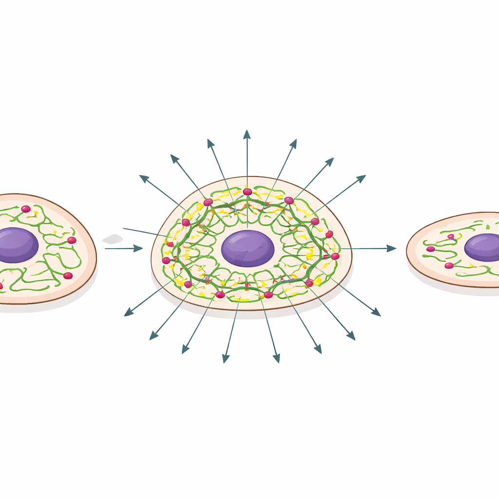
To uncover how KIF20A exerts its influence, the team searched for proteins that might physically interact with it. A network analysis highlighted CLIP1, a protein that attaches to the growing ends of tiny tubes inside the cell known as microtubules, which help shape the cell and guide its movement. Follow-up experiments confirmed that KIF20A and CLIP1 bind to each other both in engineered cells and in cervical cancer cells. When KIF20A levels were reduced, CLIP1 protein was broken down more quickly; when KIF20A was boosted, CLIP1 became more stable. In other words, KIF20A acts like a protector that keeps CLIP1 present and active on the cell’s inner tracks.
Breaking the chain to slow cancer
The final step was to test whether CLIP1 is truly the key downstream player in this process. The researchers forced cancer cells to make extra KIF20A, which predictably made them grow and migrate faster. But when they simultaneously lowered CLIP1 in these same cells, the tumor-promoting effects of KIF20A largely disappeared: growth slowed and movement declined. Patient tissues also showed higher CLIP1 levels in tumors compared with normal samples. Together, these findings support a model in which an overactive KIF20A–CLIP1 duo strengthens the internal scaffolding that cancer cells rely on to divide rapidly and invade surrounding tissue.
What this could mean for future care
For a layperson, the takeaway is that this study identifies a new “wiring diagram” inside cervical cancer cells: an overabundant motor protein, KIF20A, shields CLIP1 and helps tumor cells grow and spread. Because both proteins are much more active in cancer than in healthy tissue, they may serve as molecular flags for earlier detection or as targets for drugs designed to weaken the tumor from within. While more work is needed in larger patient groups and to clarify the exact chemical steps involved, this KIF20A–CLIP1 partnership offers a promising new angle for understanding and ultimately treating cervical cancer.
Citation: Ma, X., Xu, Z., Chen, Y. et al. KIF20A promotes cervical cancer progression by interacting with CLIP1. Sci Rep 16, 11838 (2026). https://doi.org/10.1038/s41598-026-42883-4
Keywords: cervical cancer, KIF20A, CLIP1, tumor progression, microtubule proteins