Clear Sky Science · en
Contrast enhancement for brain MRI images via genetic algorithm-based dual cut histogram equalization
Sharper Brain Scans for Earlier Answers
When doctors look for brain tumors, they often rely on MRI scans. But many of these images are low in contrast, making the difference between healthy tissue and a possible tumor frustratingly subtle. This paper introduces a new computer-based method that makes brain MRI images clearer and sharper, aiming to help radiologists and AI systems spot tumors more reliably while still keeping the overall image faithful to the original scan.
Why Fuzzy Brain Images Are a Problem
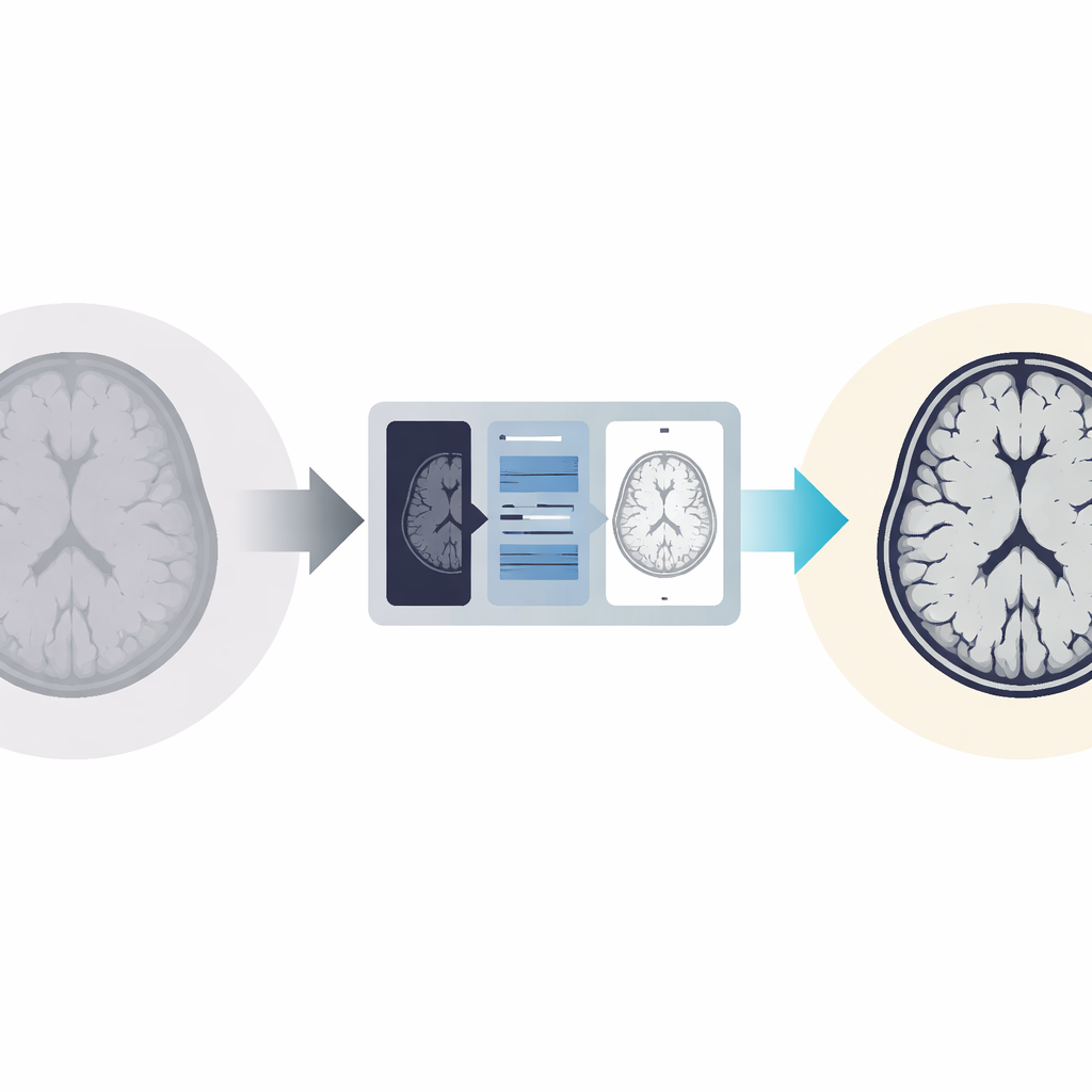
In a typical brain MRI, shades of gray represent different structures: dark areas might be background, mid-gray shows brain tissue, and brighter spots can indicate bone or a tumor. When contrast is poor, these shades blend together, hiding small or early-stage problems. This affects not only specialists reading the scans by eye, but also modern machine-learning tools that depend on clear differences in brightness. For years, researchers have used a family of tricks called histogram equalization to stretch and redistribute brightness levels across an image, making dark regions darker and bright regions brighter. While these methods can improve visibility, they often make images look harsh or unnatural, or they distort overall brightness in ways that may mislead diagnosis.
From One-Size-Fits-All to Tailored Enhancement
Traditional enhancement methods tend to treat all pixels in a rigid way. Some split the brightness range into simple halves or a few preset regions and enhance each part separately. Others, like popular “contrast-limited” approaches, divide the image into tiles and process each tile independently to avoid over-boosting noise. These strategies help, but they assume that every image fits the same pattern, which is rarely true in real-world medical data. The new technique proposed in this study takes a more flexible route. Instead of blindly following fixed rules, it analyzes how pixel brightness is actually distributed in a given brain MRI and then reshapes that distribution in a way that better separates suspected abnormal areas from normal tissue.
Cutting the Brightness Range in Three Smart Pieces
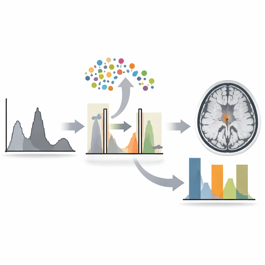
The heart of the method is called Dual-Cut Histogram Equalization. Think of the image’s brightness values as a mountain range plotted on a graph, with peaks where many pixels share similar intensity. In brain MRIs, one big cluster usually represents ordinary brain tissue, while a smaller cluster at higher brightness may contain skull or tumor regions. The authors introduce two cutting points along this brightness range, dividing it into three zones: darker background, mid-level normal tissue, and brighter structures. The middle zone, which largely corresponds to normal brain, is deliberately suppressed so it appears darker, while the darker and brighter zones are stretched in different ways. This asymmetric treatment makes potential tumors stand out more clearly against a subdued background, but still aims to avoid the harsh, artificial look often produced by simpler methods.
Letting Evolution Pick the Best Settings
Choosing exactly where to place those two cuts is critical. If they are set poorly, important details may disappear or the image may become too distorted. Instead of hand-tuning these values, the researchers use a genetic algorithm, a kind of search strategy inspired by evolution. They start with many random pairs of cut points and repeatedly “evolve” them, keeping those that produce better images and combining them into new candidates. The quality of each candidate is judged using several numerical measures of contrast and structural similarity, designed to balance visibility of fine detail against faithfulness to the original scan. This automated process adapts the enhancement to each individual MRI, rather than relying on a single recipe for all images.
Clearer Views Without Losing the Big Picture
To test their approach, the authors applied it to 1,838 brain MRI images drawn from three public datasets and compared the results with several well-known enhancement techniques. Their method consistently produced higher scores on measures that reward strong but well-distributed contrast, while still keeping image structure reasonably close to the original, as shown by standard similarity and brightness-preservation scores. Visual examples reveal tumors that stand out more distinctly after processing. In plain terms, the new method makes brain scans sharper where it matters most—around suspicious regions—without radically rewriting the picture. If adopted in clinical workflows or as a preprocessing step for AI systems, this approach could help catch brain tumors more reliably, especially in challenging low-contrast scans.
Citation: Bukhori, I., Sim, K.S., Gan, K.B. et al. Contrast enhancement for brain MRI images via genetic algorithm-based dual cut histogram equalization. Sci Rep 16, 13834 (2026). https://doi.org/10.1038/s41598-026-42868-3
Keywords: brain MRI, tumor detection, image contrast, genetic algorithm, medical imaging