Clear Sky Science · en
Deep learning–based basilar artery wall and lumen segmentation from 1-mm MR vessel wall imaging
Why this brain artery matters
Strokes in the back part of the brain can strike without clear warning signs and are notoriously hard to diagnose. At the heart of this vulnerable region lies the basilar artery, a key blood vessel running up the brainstem. This study shows how high‑resolution MRI scans, paired with artificial intelligence, can map the shape of this artery in detail, potentially helping doctors spot dangerous narrowings or bulges earlier and with less guesswork.
Looking closely at a hidden blood vessel
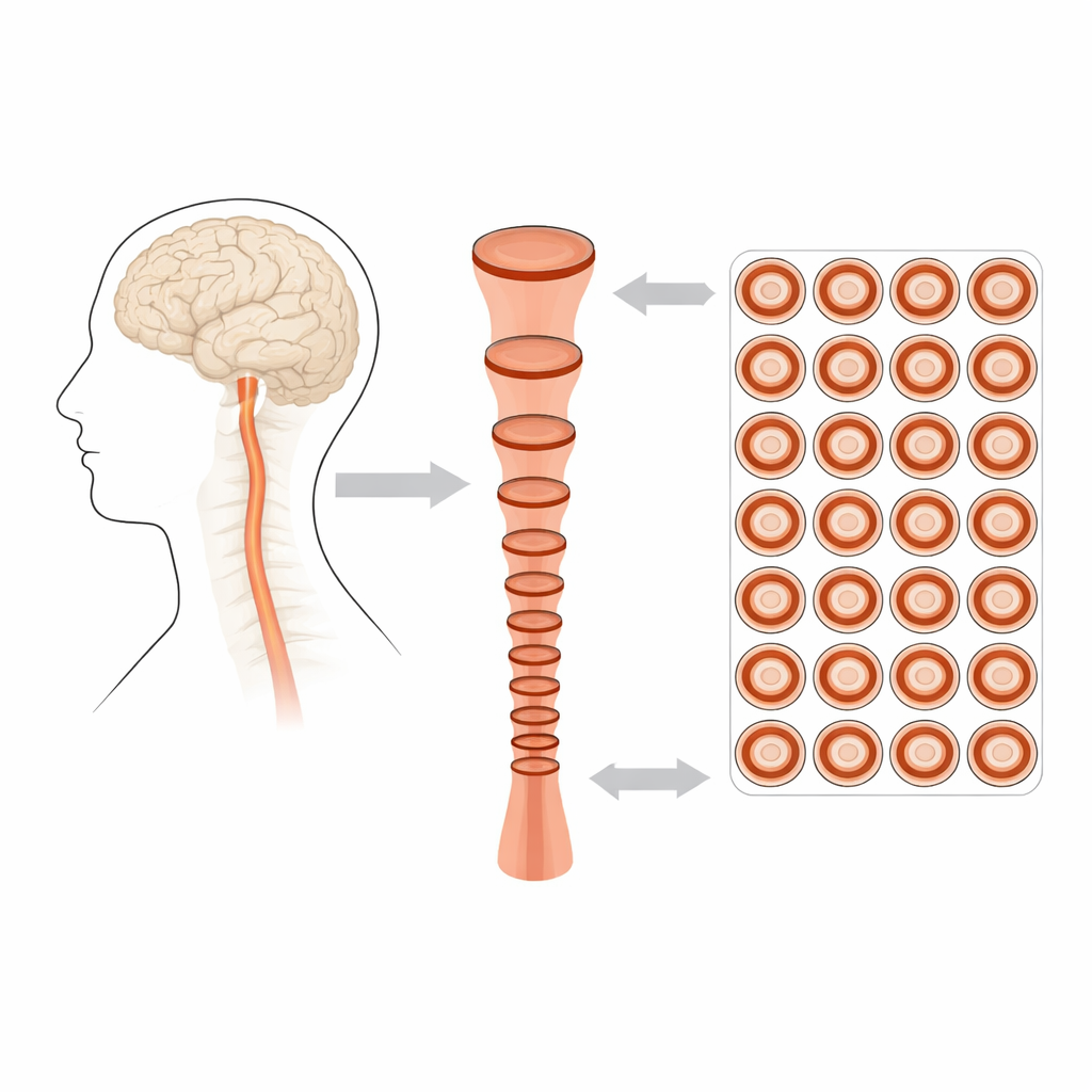
The basilar artery sits deep at the base of the brain, where traditional imaging has struggled to show more than a rough outline of its inner channel. Its wall is thin, its course is curved, and nearby structures can blur the view. Yet subtle changes in its diameter or wall thickness can signal a higher risk of stroke. The researchers used a specialized MRI approach called vessel wall imaging, which produces thin, 1‑millimeter slices with strong contrast between blood, vessel wall, and surrounding tissue. They collected scans from 36 patients being evaluated for atherosclerosis, creating hundreds of cross‑sectional images along the full length of each person’s basilar artery.
Teaching a computer to trace the artery
Manually outlining the artery wall on every slice is slow and can vary from one expert to another. To tackle this, the team adapted a powerful deep learning model, Mask R‑CNN, originally developed for general object detection. First, they manually marked the outer edge of the artery on resampled cross‑section images, then used a mathematical method to estimate the inner edge of the wall. These examples taught the model to recognize the artery as a ring‑shaped structure. After training on more than a thousand labeled slices and validating on additional scans, the algorithm could automatically detect and segment the artery in new images with a high degree of overlap compared to human‑made masks.
What the measurements revealed
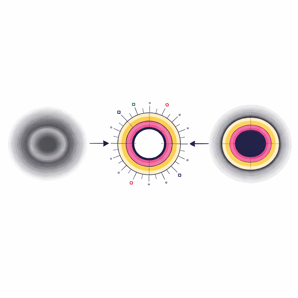
With the artery outlined automatically, the researchers could measure its inner channel—the lumen—along its full length. They found that, in most people, the basilar artery tapers gradually: it is widest at its origin and becomes significantly narrower toward its far end. On average, the lumen diameter shrank from just over 3.1 millimeters near the start to about 2.7 millimeters near the tip. When they compared the computer’s diameter measurements with those of an experienced neuroradiologist, the agreement ranged from moderate to good, suggesting the model can reliably capture overall artery size. The computer also agreed very closely with the mathematical method used to define the inner wall, reinforcing its consistency.
Limits of seeing a very thin wall
Measuring wall thickness proved much harder. The true wall of the basilar artery is thinner than the size of a single MRI pixel in this scan protocol. As a result, both humans and the AI tended to overestimate how thick the wall was, and their measurements did not match well. In areas where the artery lies close to the brainstem or where tiny branches peel off, the model sometimes misjudged the outer boundary. Even so, the combination of centerline‑based resampling and AI segmentation made it easier to visualize bright spots and bulges in the wall that may correspond to atherosclerotic plaques, as well as unusual patterns such as a widening of the distal segment where most arteries normally narrow.
What this could mean for stroke care
This work shows that a practical, 1‑millimeter MRI protocol, paired with a tailored deep learning model, can reliably capture the overall shape and diameter of the basilar artery in living patients. Understanding that a gentle tapering is normal helps doctors distinguish natural variation from truly abnormal widening or narrowing. While this approach is not yet precise enough to measure the artery wall itself in absolute terms, it can highlight suspicious changes in lumen size and shape that may warrant closer follow‑up or higher‑resolution scans. With further refinement and automation, such tools could support safer planning for brain procedures and offer a noninvasive way to monitor disease in one of the brain’s most critical and hardest‑to‑see arteries.
Citation: Tsou, CH., Liu, HM. & Huang, A. Deep learning–based basilar artery wall and lumen segmentation from 1-mm MR vessel wall imaging. Sci Rep 16, 11903 (2026). https://doi.org/10.1038/s41598-026-42847-8
Keywords: basilar artery, stroke imaging, deep learning, vessel wall MRI, atherosclerosis