Clear Sky Science · en
Anti-inflammatory treatment confirms rsfMRI and TSPO PET as biomarkers of functional connectivity and neuroinflammation in rat contusion spinal cord injuries
Why this research matters for spinal cord injuries
When the spinal cord is damaged in an accident, the initial blow is only the beginning. A wave of swelling, inflammation, and cell death unfolds over days and weeks, often turning a partial injury into a life-changing disability. Doctors urgently need ways to see this hidden process inside the cord and to tell whether a treatment is truly helping. This study in rats shows that two advanced scanning methods can act like "health meters" for the injured spinal cord, tracking both nerve network health and inflammation as a proven drug treatment does its work.
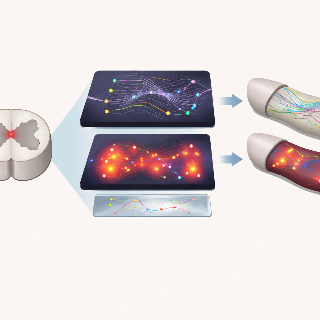
Looking inside the injured spinal cord without surgery
The researchers focused on two noninvasive imaging tools. The first is resting-state functional MRI, which tracks tiny changes in blood oxygen to infer how strongly different regions of the nervous system are working together. In the spinal cord, this reveals how well the gray matter "horns" on each side of the cord stay in sync. The second is a type of PET scan that uses a radioactive tracer designed to stick to a protein found at higher levels when immune cells in the nervous system are activated, a signature of inflammation. Together, these techniques offer a way to see both the communication networks and the inflammatory response in the same injured tissue over time.
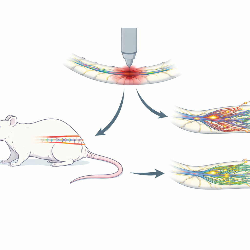
Testing a known protective drug in a rat injury model
To put these imaging methods to the test, the team used a well-established rat model of spinal cord injury. Sixteen male rats received a moderate bruise-like impact to the lower spinal cord, similar in character to many human contusion injuries. Half the animals were given riluzole, a drug already known to protect nervous tissue and help motor recovery, while the other half received an inactive solution. Over the following weeks, the rats underwent repeated MRI and PET scans and a battery of movement and sensation tests that measured walking ability, responses to touch, and sensitivity to heat. This design allowed the scientists to ask whether scan changes matched both the injury’s evolution and the animals’ functional recovery.
Tracking nerve network strength after injury
The MRI findings showed that the drug-treated animals preserved spinal cord communication better than untreated rats, especially early after injury. In regions just below the injury site, the connection strength between the back portions of the gray matter on each side of the cord was clearly higher in riluzole-treated rats during the first week. In both groups, many connections gradually weakened over four weeks, reflecting continuing deterioration of local circuits. Yet the pattern of connectivity over time closely tracked changes in movement and sensory tests: when connections were stronger, animals tended to walk better and showed more normal responses to touch and heat. Interestingly, a separate measure of overall signal strength did not differ between groups, suggesting that what mattered was how well regions stayed coordinated, not just how active they were.
Imaging inflammation and tissue response
The PET scans confirmed that spinal cord injury triggers a surge of inflammatory activity at the damage site. Rats with real injuries showed higher uptake of the inflammation-targeting tracer than sham-operated rats who underwent surgery without cord damage. However, PET could not clearly distinguish between riluzole-treated and untreated injured animals, even though post-mortem tissue staining showed fewer activated immune cells in treated cords. This suggests that while PET is sensitive to the presence of injury-related inflammation, it may be less able to pick up moderate reductions produced by this particular treatment under the conditions tested. The combination of imaging and tissue analysis nevertheless reinforced that riluzole reduced inflammation and that MRI connectivity measures captured meaningful functional differences.
What this means for future treatments
Taken together, the results show that advanced MRI and PET scans can serve as informative biomarkers—objective readouts—of what is happening inside an injured spinal cord over time. Resting-state MRI, in particular, captured early protection of spinal networks by riluzole and closely mirrored changes in movement and sensation. PET scans reliably detected injury-related inflammation and, combined with tissue studies, confirmed that the drug has anti-inflammatory effects even when subtle changes are hard to see. By providing noninvasive ways to gauge injury severity, monitor progression, and evaluate whether a therapy is working, these imaging tools could speed the development and testing of new spinal cord treatments and may ultimately help clinicians personalize care for people living with spinal cord injuries.
Citation: Mu, C., Reed, J.L., Wang, F. et al. Anti-inflammatory treatment confirms rsfMRI and TSPO PET as biomarkers of functional connectivity and neuroinflammation in rat contusion spinal cord injuries. Sci Rep 16, 14066 (2026). https://doi.org/10.1038/s41598-026-42844-x
Keywords: spinal cord injury, neuroinflammation, functional MRI, PET imaging, riluzole