Clear Sky Science · en
Anterior papillary muscle strain predicts significant left anterior descending coronary artery stenosis in patients undergoing angiography
Why this heart study matters
Many people worry about clogged heart arteries but hope to avoid invasive tests that involve threading a tube into the heart. This study asked whether a tiny structure inside the heart—the anterior papillary muscle—could act as an early warning sign of serious narrowing in a major heart artery, using only an ultrasound scan from outside the chest. If this “hidden muscle signal” worked, it might help doctors decide who truly needs an angiogram and who can be safely spared the procedure.
A closer look at a key heart artery
The researchers focused on the left anterior descending artery, often nicknamed the “widow‑maker” because a severe blockage in its early segment can have grave consequences. They studied 130 adults who were already scheduled for coronary angiography, the dye‑and‑X‑ray test that directly shows artery narrowings. Half of the participants had a significant tightening (70% or more) in the early part of this artery, while the other half had arteries that appeared normal. This design allowed the team to compare subtle heart‑muscle behavior in patients with and without a clearly dangerous blockage.
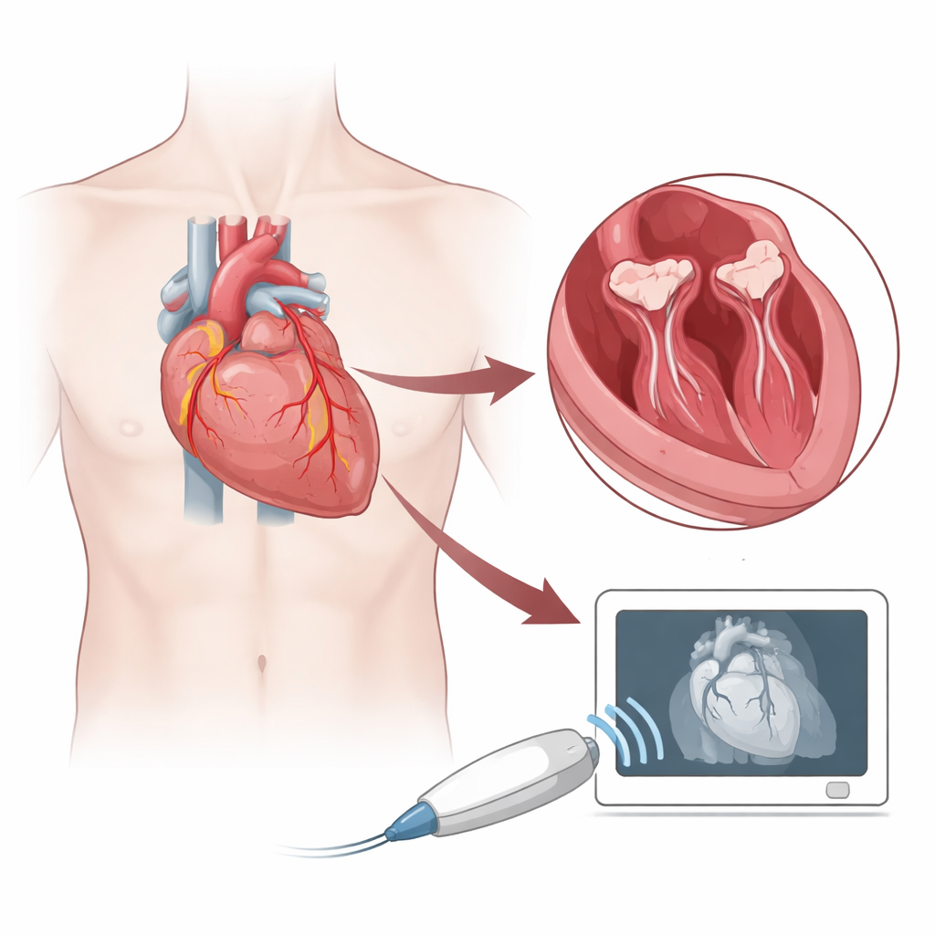
The tiny muscles behind a big heartbeat
Inside the main pumping chamber of the heart, small finger‑like muscles—papillary muscles—help the mitral valve open and close cleanly with each beat. The anterolateral papillary muscle has blood supply from branches of both the left anterior descending and another artery, which may make it somewhat resilient but also closely linked to disease in that crucial front artery. Using advanced echocardiography, a sophisticated form of ultrasound, the team tracked how much this papillary muscle shortened and stretched during each heartbeat, a measure called longitudinal strain. They also recorded more familiar readings such as overall pumping strength, filling pressures, and how well the whole heart muscle lengthened and shortened.
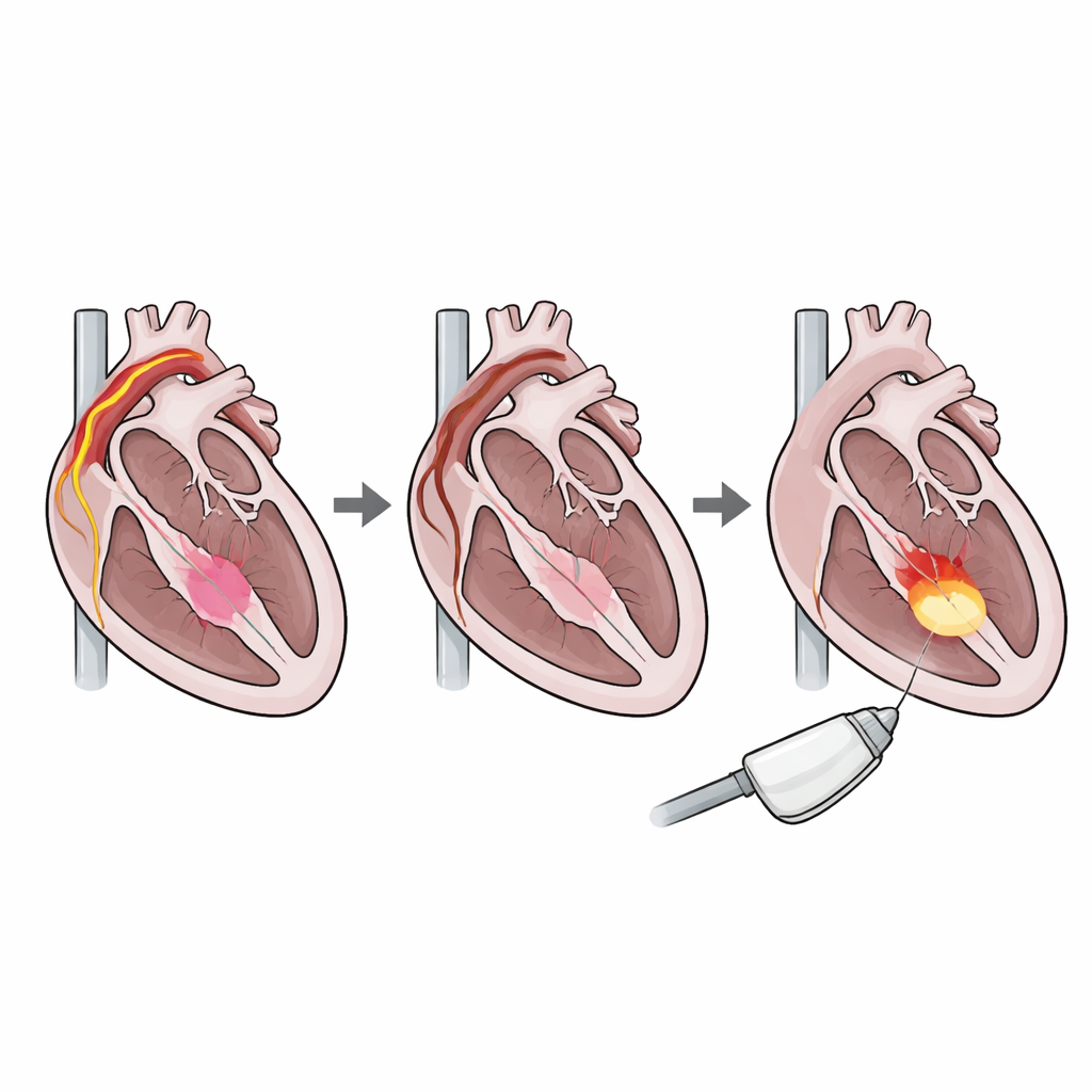
What the scans revealed
When the numbers were first analyzed by themselves, the papillary muscle’s strain looked promising. Patients with a tight narrowing in the left anterior descending artery tended to have less vigorous shortening of the anterolateral papillary muscle. In statistical terms, each step toward weaker strain slightly increased the odds of having a serious blockage. Even after accounting for obvious wall motion problems seen on routine ultrasound images, this relationship still held. This suggested that papillary muscle strain was indeed sensing the impact of reduced blood flow in that artery.
Why the signal may not stand alone
However, the picture changed when the researchers adjusted for a broader set of heart performance measures, including overall ejection fraction, a key filling‑pressure ratio, and global strain of the entire left ventricle. Once these were considered together, papillary muscle strain no longer added clear, independent information. The small muscle’s behavior was so closely tied to how the rest of the heart was functioning that its unique contribution faded in the math. Careful simulations also showed that the study, while thoughtfully designed, might not have included enough people to reliably detect a modest independent effect after all these adjustments.
What this means for patients
For now, the findings suggest that measuring strain in this single papillary muscle, on its own, is not ready to replace or dramatically reshape current strategies for deciding who needs an angiogram. It does appear to mirror disease in an important artery, but much of that information may already be captured by existing ultrasound measures of overall heart function. Future, larger studies may test whether papillary muscle strain can still add value when combined smartly with other imaging results and clinical risk factors. In everyday terms, this tiny muscle might yet become part of a more refined, noninvasive “fingerprint” of coronary disease, but it is not a stand‑alone screening test today.
Citation: Bagheri, A., Khani, M., Bozorgi, S.J. et al. Anterior papillary muscle strain predicts significant left anterior descending coronary artery stenosis in patients undergoing angiography. Sci Rep 16, 13446 (2026). https://doi.org/10.1038/s41598-026-42747-x
Keywords: coronary artery disease, papillary muscle, echocardiography, heart strain imaging, LAD stenosis