Clear Sky Science · en
Profiling extracellular matrix-driven heterogeneity of single cell migration and morphology
Why the cell’s neighborhood matters
Inside our bodies, cells are constantly on the move—healing wounds, shaping developing tissues, and, in unfortunate cases, spreading cancer. But cells don’t travel through empty space. They crawl across a molecular “floor” called the extracellular matrix, a web of proteins that surrounds and supports them. This study asks a deceptively simple question: if you change that floor, do cancer cells move and look different—and can modern image analysis help us read those changes in a quantitative, unbiased way?
Three different cellular playing fields
The researchers focused on HeLa cells, a widely used cancer cell line, and placed them on dishes coated with three common matrix proteins: laminin and two types of collagen. Laminin often lines natural barriers in the body, while collagens form tough fibers that give tissues strength. Using time-lapse microscopy over 12 hours, the team recorded thousands of cells as they crawled across these different surfaces. Automated tools based on modern computer vision first detected and tracked individual cells, then measured how far and how fast they moved, how often they paused or turned, and how much area each cell covered.
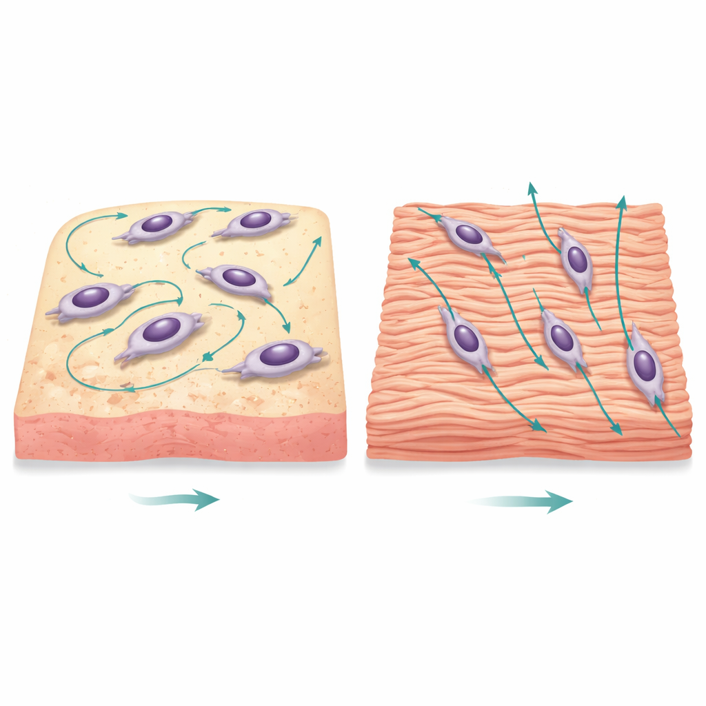
Different floors, different ways of moving
At first glance, the tracks of cells on laminin looked more confined than those on collagen, as if they were pacing in place. Yet when the team quantified the movements, a more nuanced picture emerged. Cells on laminin actually traveled slightly longer total distances, but their net progress from start to finish was smaller. They frequently changed direction, had larger turning angles, and showed lower “persistence,” meaning they did not stick to a straight path for long. In contrast, cells on both collagen types tended to move more directly, covering similar distances overall but ending up farther from where they started. Statistical measures confirmed that the two collagen conditions behaved much like each other, but distinctly from laminin.
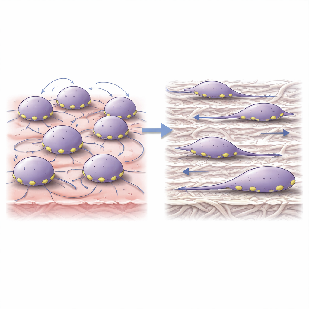
Shape and structure tell an added story
From the same movies, the authors extracted each cell’s outline to characterize its shape. On laminin, cells spread out more, covering larger areas with less elongated, more compact forms. On collagen, cells appeared thinner and more stretched. To capture all of the motion and shape information at once, the researchers used a standard statistical tool that condenses many measurements into a few combined “axes” of variation. This analysis clearly separated laminin-grown cells from collagen-grown cells, especially when focusing on movement-related traits like turning, pausing, and displacement, while the differences in overall shape were present but subtler.
How cells grip and talk to each other
Numbers alone do not explain why cells behave differently, so the team turned to cell biology. They examined how often cells touched their neighbors and how their internal support structures were organized. On laminin, cells formed more frequent and longer-lasting contacts with each other, often via slender projections reaching out like feelers. The sites where cells anchor to the matrix—tiny “feet” called focal adhesions—also differed: on laminin, cells had many more of these adhesions, but each one was smaller; on collagen, adhesions were fewer but larger. Previous work suggests that small, rapidly turning over adhesions favor agile, exploratory movement, whereas large, stable adhesions support slower, more directed motion. The patterns observed here fit that picture and help explain the distinct migration styles.
A framework for reading cell behavior from images
Taken together, this work shows that swapping one matrix protein for another can shift cancer cells from straight-ahead travel to a more searching, flexible mode of movement with richer cell–cell interactions. By combining automated image analysis with transparent statistical methods, the study links those behavioral changes to concrete biological features, such as how cells spread, how they connect to neighbors, and how they grip their surroundings. Because the approach is scalable and reproducible, it could be extended to other cell types, more complex tissue-like environments, and even drug testing. For non-specialists, the key message is that the “ground” cells walk on is not just passive support—it actively guides how they move, interact, and potentially spread disease, and new computational tools are making these hidden influences visible and measurable.
Citation: Shin, E., Han, J., Jung, A. et al. Profiling extracellular matrix-driven heterogeneity of single cell migration and morphology. Sci Rep 16, 12609 (2026). https://doi.org/10.1038/s41598-026-42530-y
Keywords: cell migration, extracellular matrix, cancer cell behavior, cell mechanics, image-based analysis