Clear Sky Science · en
Yolov8n based on dynamic serpentine convolution and multi-feature attention for MRI brain cranial tumor segmentation
Why this matters for patients and doctors
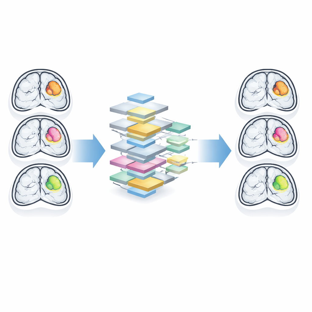
Brain tumors are life‑threatening not only because of how fast they grow, but also because they sit in the body’s most delicate organ. Before surgery or radiation, doctors must know exactly where a tumor begins and ends on MRI scans. Today, this outlining is often done by hand and can be slow, tiring, and imperfect. This paper introduces a new artificial‑intelligence approach that aims to trace brain tumors automatically, quickly, and with sharper edge detail, potentially giving neurosurgeons clearer maps to guide treatment.
Seeing tumors clearly in a noisy picture
Magnetic resonance imaging (MRI) is the main tool for spotting brain tumors and planning operations. Yet brain tumors come in many forms—some are round and well defined, others are stringy, patchy, or intertwined with normal tissue. Their borders can be fuzzy, especially in early stages, making it hard even for experts to draw precise outlines slice by slice. Traditional computer methods either rely on hand‑crafted rules from mathematics or on standard deep‑learning models that blur details as they repeatedly compress and re‑expand the image. Both approaches struggle most where precision matters most: at the tumor’s edge.
A faster detector tuned for medicine
The authors build on a family of AI models known as YOLO, originally designed for real‑time object detection in photos and video. YOLO is fast and efficient—qualities that are valuable in busy hospitals where treatment decisions must be made quickly. However, the basic YOLO models were created for everyday scenes, not for subtle medical nuances. To adapt YOLOv8n (a compact version of the model) to brain MRI scans, the researchers re‑engineer its internal blocks so that it pays special attention to fine structures and irregular shapes rather than just broad objects.
A snake‑like lens that follows tumor edges
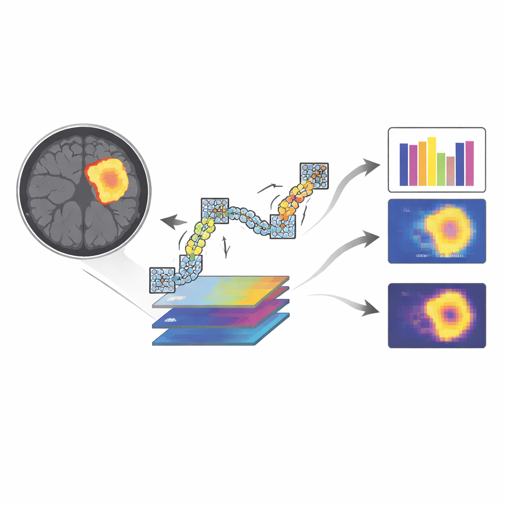
The first major innovation is what the authors call dynamic serpentine convolution. Standard AI “lenses” look at image patches on a rigid square grid. That works well for cars and street signs, but not for tumors that stretch and bend through brain tissue in winding, tube‑like patterns. Dynamic serpentine convolution, by contrast, lets the lens slide along flexible, snake‑shaped paths that adapt to local structure. As the model scans an MRI slice, its sampling points are gently steered to follow continuous curves instead of fixed boxes, making it naturally better at tracing complex contours without losing track of where it is.
Combining many views and focusing on what matters
To further boost accuracy, the model does not rely on a single pass of this snake‑like scanning. It applies several such passes to the same image region and then fuses the resulting feature maps into a richer, combined description—a strategy the authors call multi‑feature fusion. On top of this, they add a dual‑attention mechanism. One part of this mechanism works across channels, deciding which kinds of features (such as texture or brightness patterns) deserve more weight. The other part works across space, highlighting the most informative locations in the image. Together, these attention layers act like a radiologist who both knows which cues usually signal a tumor and zooms in on suspicious spots instead of treating every pixel equally.
How well does the new method work?
The team trained and tested their improved model, called DMA‑YOLOv8n, on a public dataset of 1,300 brain MRI images, and also checked how well it transfers to another well‑known brain tumor collection (BraTS2021). They judged performance using standard measures of how often the automatic outlines match expert‑drawn ones. Compared with the original YOLOv8n and several other leading systems, including popular U‑Net variants used in hospitals and research labs, DMA‑YOLOv8n achieved the best balance of accuracy and efficiency. It reached higher precision and recall scores, indicating that it both missed fewer tumor pixels and mislabeled fewer healthy pixels as tumor, while keeping computation costs modest enough for practical use.
What this means for future care
From a layperson’s perspective, the key takeaway is that this new AI method can draw tumor borders on brain scans more sharply and more reliably than many existing tools, without slowing down care. By following the natural, winding patterns of tumor growth and by learning to focus on the most telling details, DMA‑YOLOv8n produces outlines that closely match expert opinion. If integrated into clinical workflows, such systems could help doctors plan surgery and radiation with greater confidence, shorten the time from diagnosis to treatment, and potentially improve outcomes for patients facing brain tumors.
Citation: Hang, Y., Zhang, Q., Li, L. et al. Yolov8n based on dynamic serpentine convolution and multi-feature attention for MRI brain cranial tumor segmentation. Sci Rep 16, 12008 (2026). https://doi.org/10.1038/s41598-026-42502-2
Keywords: brain tumor MRI, medical image segmentation, deep learning, YOLOv8, clinical decision support