Clear Sky Science · en
An optimized EfficientNetB0 framework with CLAHE-based preprocessing for accurate multi-class chest X-ray classification
Smarter Help from Chest X‑Rays
Chest X‑rays are among the most common medical tests in the world, yet reading them correctly—especially when several lung or heart problems appear at once—is far from simple. This study describes a new computer system that helps doctors spot multiple chest diseases in a single X‑ray more accurately, using a carefully tuned artificial intelligence (AI) model and image‑enhancement steps that make subtle details easier to see.
Why Chest Images Are Hard to Read
On a chest X‑ray, many layers of organs and tissues overlap: ribs, lungs, heart and blood vessels all pile into the same flat picture. Experienced radiologists learn to untangle this visual puzzle, but even they can struggle when several conditions occur together, such as fluid around the lungs, infection and an enlarged heart. Earlier computer‑aided tools often simplified this reality, treating each picture as if it showed just one problem or none at all. That makes them less useful in real clinics, where patients frequently have more than one disease at the same time.
Building a Cleaner View of the Lungs
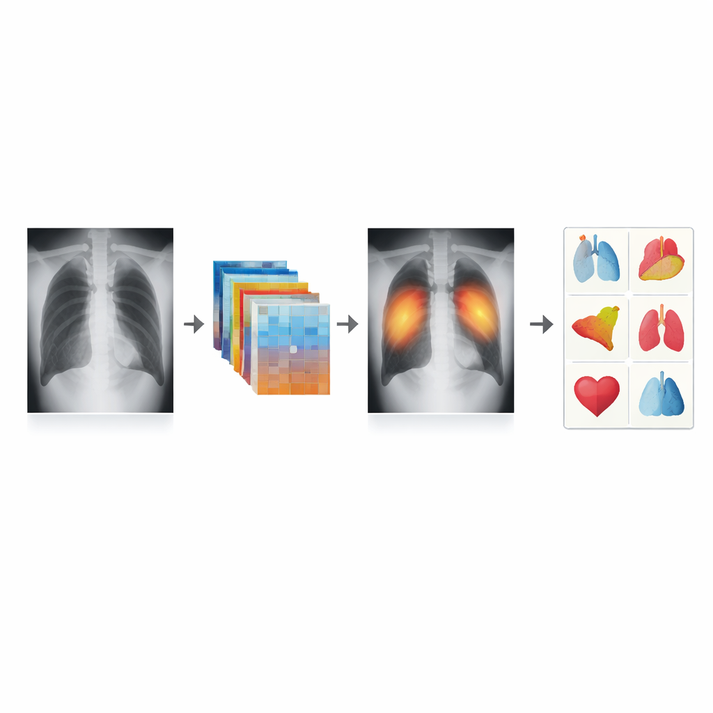
The researchers began with a large public collection of more than 100,000 anonymized chest X‑rays from the U.S. National Institutes of Health. They focused on five key findings that matter in everyday care: normal exams, pneumonia, collapsed lung (pneumothorax), fluid around the lung (effusion) and an enlarged heart (cardiomegaly). Before letting the AI learn from the images, they passed every X‑ray through a standardized cleaning pipeline. The images were kept in gray scale, their contrast was gently boosted using a method called CLAHE to reveal faint patterns in the lungs, brightness was adjusted for consistency and all pictures were resized to the same dimensions. The team also balanced rare and common findings by carefully repeating some examples and trimming others, so that the model would not ignore uncommon but important diseases.
Training an AI That Respects Real‑World Complexity
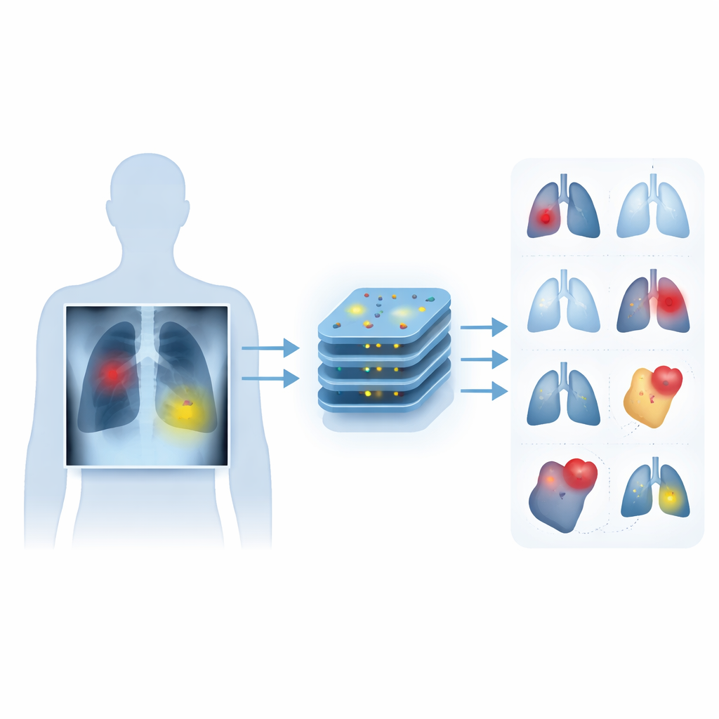
At the heart of the system lies an EfficientNetB0 network, a modern deep‑learning design known for squeezing strong performance out of relatively small, fast models. The authors customized this network for the mixed reality of chest imaging: instead of forcing a single yes‑or‑no answer, the system outputs a separate probability for each of the five findings, allowing it to mark several problems in one image. They added an attention‑like component that helps the model emphasize the most informative patterns, and used a special training rule that focuses learning on the hardest, easily missed cases. Training was done in two stages—first adjusting only the final layers, then fine‑tuning the entire network—using rigorous cross‑validation to check that results were stable and not due to chance splits in the data.
How Well the System Performs
To see whether their design truly helped, the team compared EfficientNetB0 with two popular alternatives, DenseNet121 and MobileNetV2, under identical conditions. All three models achieved similar overall accuracy, but the optimized EfficientNetB0 stood out where it counts most in medicine: it was better at separating sick from healthy cases and at avoiding missed diagnoses. Across all five findings, it reached a high average score for distinguishing disease from non‑disease, and it was especially strong for pneumonia and an enlarged heart. Even for the trickier collapsed lung and fluid cases, where X‑ray signs can be subtle and overlap with other problems, the model maintained solid detection ability while keeping consistency across repeated tests.
What This Means for Patients and Clinicians
The study shows that paying attention to the full workflow—from how X‑rays are cleaned and balanced to how the AI is trained—can produce a tool that better mirrors the messy reality of hospital imaging. Rather than aiming to replace radiologists, the proposed system is designed to act as a second set of eyes, flagging multiple possible problems on a single chest X‑ray and drawing attention to cases that deserve closer human review. With its strong results on a large public dataset and a design that can be adapted to other collections, this approach moves computer‑aided diagnosis a step closer to everyday use, where faster and more reliable readings of chest X‑rays could help patients receive timely, appropriate care.
Citation: Hegazy, N.Y., Sawah, M.S. An optimized EfficientNetB0 framework with CLAHE-based preprocessing for accurate multi-class chest X-ray classification. Sci Rep 16, 10811 (2026). https://doi.org/10.1038/s41598-026-42492-1
Keywords: chest X-ray AI, lung disease detection, deep learning radiology, medical image preprocessing, multi-label classification