Clear Sky Science · en
Magnification-independent breast cancer diagnosis using a GWO-enhanced vision transformer with multi-stage stain normalization
Why this research matters to patients and doctors
Breast cancer is now the most commonly diagnosed cancer worldwide, and the microscopic examination of tissue samples remains the gold standard for confirming the disease. Yet reading these slides is time‑consuming, can vary from one expert to another, and becomes even harder when images are taken at different zoom levels or stained slightly differently in the lab. This study explores how a new generation of artificial intelligence (AI) tools can help pathologists spot cancer more consistently by teaching computers to see patterns in breast tissue slides, no matter how they were captured.
Bringing order to messy microscope pictures
In real hospitals and labs, breast tissue slides rarely look exactly the same. Differences in dyes, lighting, and scanner settings can make colors shift from deep purple to pale pink, even when the underlying tissue is similar. The authors tackle this problem first, before any AI is trained. They design a four‑step cleaning pipeline that adjusts contrast, lines up overall brightness, corrects uneven lighting, and finally standardizes how the common hematoxylin–eosin stain appears. Working on more than 7,900 images from a public dataset called BreakHis, this process makes cell structures clearer and colors more consistent across samples and across four standard microscope magnifications. 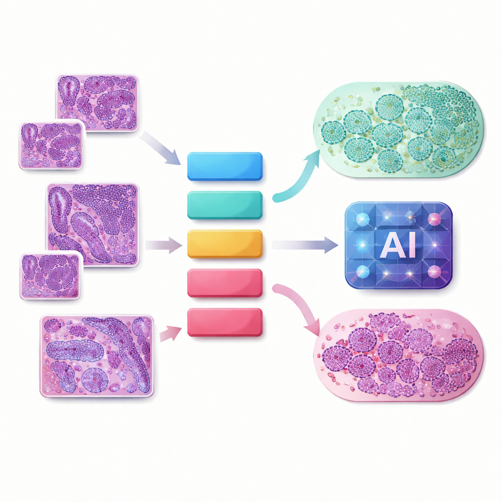
Balancing the scales between healthy and cancerous samples
A second practical issue is imbalance: the dataset contains far more cancerous images than benign ones. If left unchecked, an AI model can simply “learn” to call everything cancer and still score well on paper, while failing patients in practice. To avoid this, the researchers artificially expand the under‑represented benign class using carefully chosen transformations that mimic real‑world variation. They flip images, rotate them, slightly change brightness and color, and add gentle blur—all without destroying the fine cellular details that matter for diagnosis. After this step, benign and malignant examples are evenly matched at each magnification level, giving the model a fair chance to learn both categories.
Teaching a transformer to read tissue slides
Instead of using traditional convolutional neural networks, the team adopts a Vision Transformer—a design originally invented for language processing and later adapted to images. Rather than scanning the picture with small filters, the transformer chops each slide into many small patches and learns how these pieces relate to one another across the entire image. This global perspective is valuable for histopathology, where the shape and arrangement of cells and tissue patterns can signal whether a tumor is harmless or aggressive. The model is trained separately for each zoom level (40×, 100×, 200×, 400×), and performance is checked with standard measures such as accuracy, precision, recall, confusion matrices, and cross‑validation to ensure that results are stable and not a fluke of one lucky train–test split.
Letting a nature‑inspired optimizer fine‑tune the AI
Vision Transformers have many “knobs” that strongly influence how well they work: how deep the network should be, how many attention heads to use, how large the internal representations are, how much dropout to apply, and what learning rate to choose. Rather than tuning these by trial and error, the authors turn to a meta‑heuristic called the Grey Wolf Optimizer. Inspired by how wolves coordinate to track and encircle prey, this algorithm explores combinations of settings and gradually homes in on those that minimize validation error. Applied independently at each magnification, it discovers transformer configurations that make better use of the cleaned, balanced images. 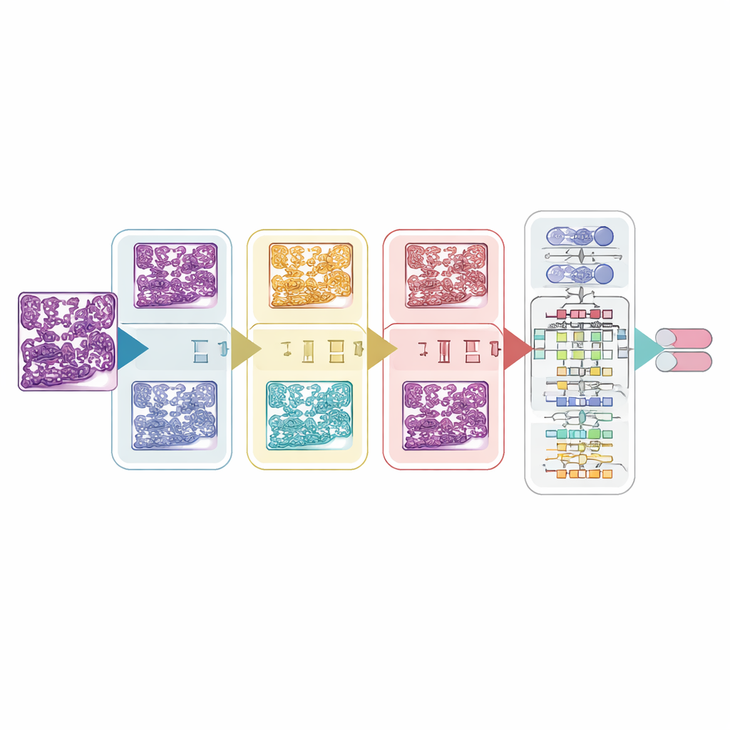
Clear gains in accuracy across all zoom levels
After optimization, the combined framework—stain‑aware preprocessing, magnification‑specific balancing, and Grey Wolf–tuned Vision Transformers—achieves strong and consistent results. Test accuracies reach about 92% at the lowest magnification and climb to 94% at the highest. Importantly, cancerous (malignant) cases are detected with slightly higher recall than benign ones, which is crucial because missing a cancer is far more harmful than flagging a benign sample for extra review. Repeating the experiments through five‑fold cross‑validation shows minimal variation, suggesting the system is learning robust cues rather than memorizing the training set.
What this could mean for future cancer care
In simple terms, the study shows that when tissue images are carefully standardized and a transformer‑based model is smartly tuned, an AI system can reliably distinguish between benign and malignant breast samples across multiple microscope zoom levels. While the work is still limited to one public dataset and a binary decision (cancer versus not), it points toward clinical tools that could help overworked pathologists by pre‑screening slides, highlighting suspicious regions, and reducing missed cancers. With broader, multi‑center validation and further refinement, such magnification‑independent AI assistants could become an integral part of digital pathology workflows, supporting faster and more consistent breast cancer diagnosis.
Citation: Fatma, T., Sahu, P.K., Choudhury, S. et al. Magnification-independent breast cancer diagnosis using a GWO-enhanced vision transformer with multi-stage stain normalization. Sci Rep 16, 11930 (2026). https://doi.org/10.1038/s41598-026-42490-3
Keywords: breast cancer histopathology, vision transformer, stain normalization, medical image classification, deep learning optimization