Clear Sky Science · en
Ocular biometry agreement among ZW-30, IOLMaster 700, and sirius in cataract patients
Why measuring the eye matters for cataract surgery
Cataract surgery has quietly become one of the most precise procedures in modern medicine. Today, surgeons do not just remove a cloudy lens; they also aim to leave patients with sharp, glasses-free vision. To do that, they rely on machines that measure the eye in great detail before surgery. This study asks a deceptively simple but important question: when three advanced devices measure the same cataract patients, do they really agree—and can doctors safely mix and match their readings?
Three different machines looking at the same eye
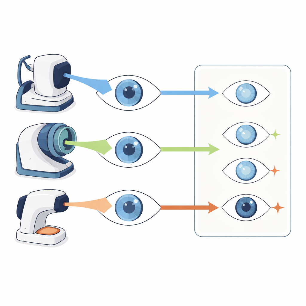
The researchers focused on three state-of-the-art instruments used before cataract surgery. Two of them, the IOLMaster 700 and the new ZW-30, scan the eye using a fast, sweeping light beam that builds up an internal picture. The third, called Sirius, combines two other imaging methods to map the front of the eye. All three devices measure how long the eye is, how thick and curved the clear front window (the cornea) is, how deep the front chamber is, and how thick the natural lens is—numbers that feed into formulas to choose the right artificial lens.
How the study was carried out
The team studied 307 people with cataracts, examining 603 eyes. Each eye was measured with all three biometers in a random order, under controlled lighting and by an experienced operator. The scientists then compared the readings in several ways: they looked at how closely the values tracked together, checked whether the average numbers matched, and examined how large the differences were from one machine to another for individual patients. They also built mathematical equations that could, in theory, convert measurements from one device into the scale of another.
Where the machines agree—and where they do not
For many basic dimensions of the eye—such as the thickness of the central cornea, the depth of the front chamber, the overall length of the eye, the thickness of the lens, and the main front-surface curvature—the three devices moved in step. On average, their numbers were very close, and the patterns across the whole group lined up well. This means that when researchers look at trends in large populations, these machines paint a similar big-picture story. However, when the scientists inspected the spread of differences for individual eyes, the range was wider than what surgeons consider safe to treat readings as interchangeable. In other words, group averages looked reassuring, but any single patient might still get meaningfully different numbers depending on which machine was used.
Hidden trouble in more subtle eye measurements
The story became more complicated for measurements that describe the eye’s fine optical details. These include the combined bending power of both the front and back of the cornea and a small alignment offset between the line of sight and the geometric center of the eye. For these subtler features, the devices showed only moderate agreement, and their average values differed in ways that could matter clinically. Even when the general trend was similar, the individual discrepancies were large. The width of the colored part of the eye and the size of the pupil were especially device-dependent, reflecting different ways the machines detect edges and capture images. Here, the authors concluded that the numbers simply cannot be swapped between devices or trusted to be equivalent.
What this means for patients and surgeons
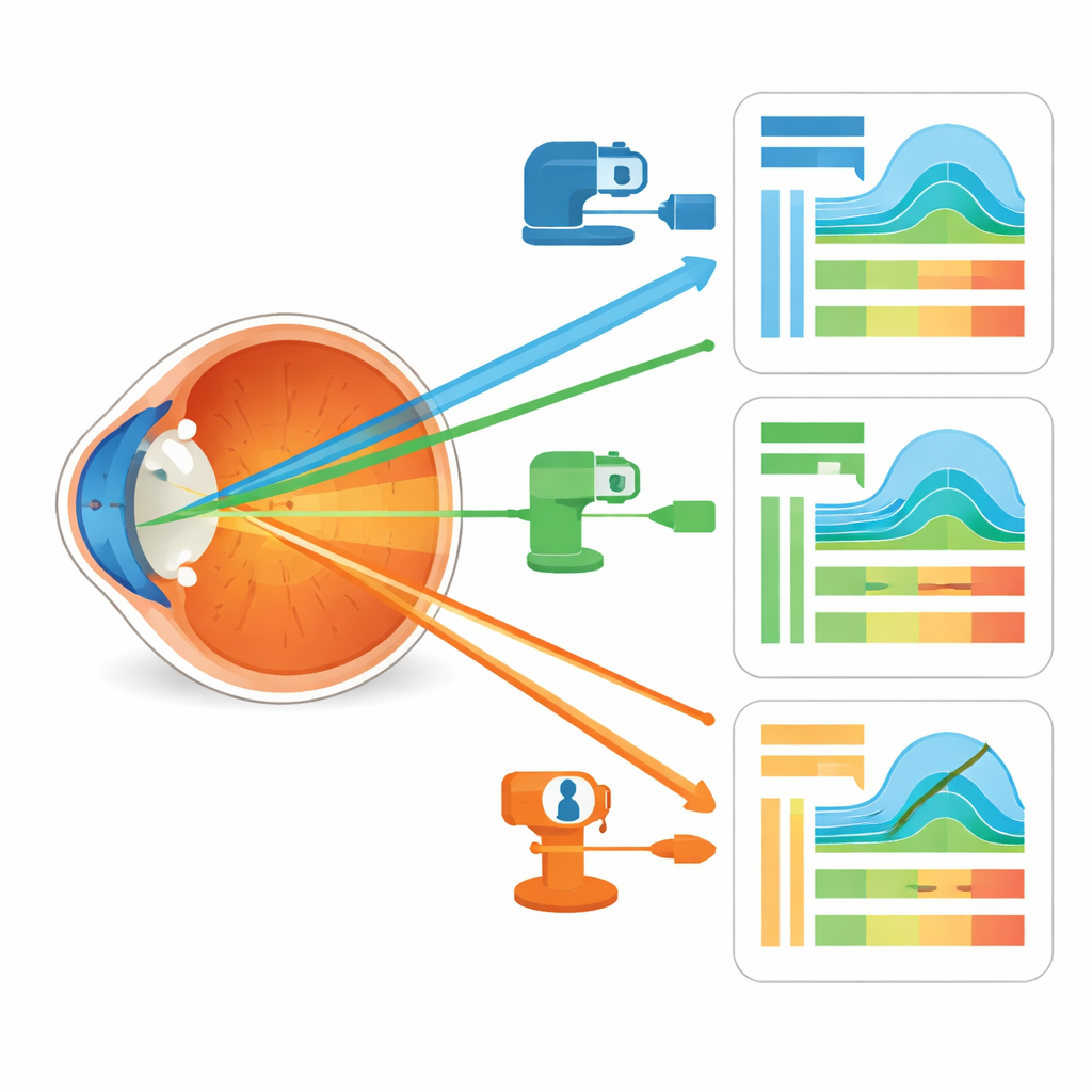
For someone facing cataract surgery, the key message is that modern measuring tools are broadly reliable but not identical. This study shows that while different biometers tell a similar story about groups of patients, they may disagree enough on a single person’s eye to influence the choice of lens power or astigmatism correction. The authors recommend that surgeons avoid mixing measurements from different machines when planning surgery and be cautious about using conversion formulas in daily practice. Future advances—such as combining multiple imaging methods in one platform or using artificial intelligence to harmonize readings—may help close these gaps and make precision vision after cataract surgery even more dependable.
Citation: Yang, J., Yang, N., Xiang, Y. et al. Ocular biometry agreement among ZW-30, IOLMaster 700, and sirius in cataract patients. Sci Rep 16, 12618 (2026). https://doi.org/10.1038/s41598-026-42451-w
Keywords: cataract surgery, ocular biometry, eye imaging devices, corneal measurements, intraocular lens planning